Brent G. Petty - Basic Electrocardiography
Here you can read online Brent G. Petty - Basic Electrocardiography full text of the book (entire story) in english for free. Download pdf and epub, get meaning, cover and reviews about this ebook. year: 2020, publisher: Springer International Publishing, genre: Children. Description of the work, (preface) as well as reviews are available. Best literature library LitArk.com created for fans of good reading and offers a wide selection of genres:
Romance novel
Science fiction
Adventure
Detective
Science
History
Home and family
Prose
Art
Politics
Computer
Non-fiction
Religion
Business
Children
Humor
Choose a favorite category and find really read worthwhile books. Enjoy immersion in the world of imagination, feel the emotions of the characters or learn something new for yourself, make an fascinating discovery.
- Book:Basic Electrocardiography
- Author:
- Publisher:Springer International Publishing
- Genre:
- Year:2020
- Rating:5 / 5
- Favourites:Add to favourites
- Your mark:
Basic Electrocardiography: summary, description and annotation
We offer to read an annotation, description, summary or preface (depends on what the author of the book "Basic Electrocardiography" wrote himself). If you haven't found the necessary information about the book — write in the comments, we will try to find it.
Brent G. Petty: author's other books
Who wrote Basic Electrocardiography? Find out the surname, the name of the author of the book and a list of all author's works by series.


Basic Electrocardiography — read online for free the complete book (whole text) full work
Below is the text of the book, divided by pages. System saving the place of the last page read, allows you to conveniently read the book "Basic Electrocardiography" online for free, without having to search again every time where you left off. Put a bookmark, and you can go to the page where you finished reading at any time.
Font size:
Interval:
Bookmark:


This Springer imprint is published by the registered company Springer Nature Switzerland AG
The registered company address is: Gewerbestrasse 11, 6330 Cham, Switzerland
To my wife, Joan, for her long-standing love and support.
To our four wonderful childrenElliott, Carter, Mason, and Hillary.
To the generation of medical students whom I have had the pleasure to teach.
This book is intended to help students of all healthcare delivery fields and all levels of training to learn the basic concepts of interpreting electrocardiograms. While originally and primarily intended to be used by third-year medical students at The Johns Hopkins University School of Medicine, this book has also been used successfully by nurse practitioners and physician assistants who work at Hopkins as well as by nurse practitioner students in training at Hopkins.
The chapters are constructed to introduce basic themes, give examples from actual patient tracings, and then provide practice by providing self-test electrocardiograms that will reinforce the concepts taught in the chapter. Additionally, the practice tracings build on the information provided in earlier chapters as well as on the features of the current one. The citations provided in the chapters are not intended to be comprehensive. In fact, some of them are nearly 100 years old and are provided for historical interest.
The electrocardiograms shown in the book are from patients, collected over many years, and recorded in one of two ways: either as single-channel sequential tracings or, more contemporaneously, as multichannel tracings recorded and displayed in at least three leads simultaneously. I believe that seeing both types of tracings will help the student become comfortable with new tracings, as well as with those that may still be recorded one lead at a time, and with tracings from old medical records that were obtained before the simultaneous lead methodology was developed.
One important principle for interpreting electrocardiograms is that nothing important occurs in only one beat in only one lead. Important findings occur in multiple beats in multiple leads, and the leads involved are a group of leads that would be expected to show the same or similar changes because of their electrical recording orientation.
An important goal of this book is to teach students the language of electrocardiograms. Like all facets of medicine, the interpretation of electrocardiograms is associated with terminology, even jargon, that has special meaning within that discipline. Becoming familiar with the terminology and the electrocardiographic appearance associated with the terms is a high priority. Clinical correlations are provided as much as applicable. On the other hand, the electrophysiological explanations for why the recordings have the appearance that they do are intentionally minimized.
While the vast majority of the tracings in this book are from my patients, I am grateful to Ms. Alicia Sasser, manager of The Johns Hopkins Hospital Heart Station, for several tracings that are included. Many thanks as well to Latasha S. Graham, my assistant, for her excellent work with the text, tables, and legends, to Anila Vijayan at Springer for her assistance with figures and the text, and to Garth Haller at Springer for his editorial assistance.
Enjoy learning about EKGs!
The heart is a remarkable organ, responsible for the coordinated pumping of blood through the pulmonary and systemic circulations. The muscular contraction of the chambers (systole), followed by a slightly longer period of relaxation (diastole), proceeds continuously, day in and day out, with precision and reliability in most circumstances. This book is an approach to the manifestations of the normal and abnormal electrical events associated with the function of the heart as a pump.
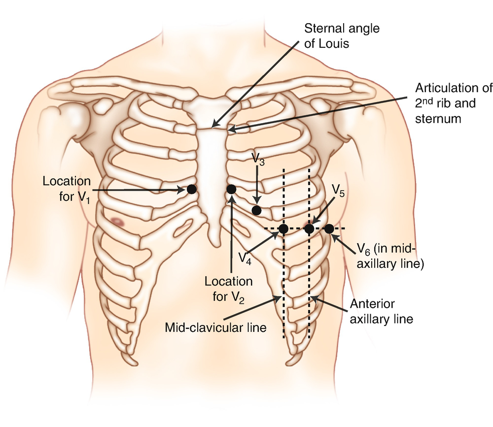
Electrocardiographic leads
Limb | Precordial |
|---|---|
I, II, III | V1, V2, V3 |
aVR, aVL, aVF | V4, V5, V6 |
Font size:
Interval:
Bookmark:
Similar books «Basic Electrocardiography»
Look at similar books to Basic Electrocardiography. We have selected literature similar in name and meaning in the hope of providing readers with more options to find new, interesting, not yet read works.
Discussion, reviews of the book Basic Electrocardiography and just readers' own opinions. Leave your comments, write what you think about the work, its meaning or the main characters. Specify what exactly you liked and what you didn't like, and why you think so.