Marc E. Grossman , Lindy P. Fox , Carrie Kovarik and Misha Rosenbach Cutaneous Manifestations of Infection in the Immunocompromised Host Second Edition 10.1007/978-1-4419-1578-8_1 Springer Science+Business Media, LLC 2012
1. Subcutaneous and Deep Mycoses
Aspergillosis
Invasive aspergillosis is the most common opportunistic fungal infection in the hematopoetic stem cell transplant patient. Prior to the regular use of antifungal prophylaxis, invasive fungal infections caused by Candida were the most common infection, followed by infection by Aspergillus .
Invasive aspergillosis occurs in the settings of severe or prolonged neutropenia due to cytotoxic therapy for leukemia or lymphoma, high-dose systemic corticosteroids for transplants or collagen vascular disease, a functional neutrophil defect, long-term immunosuppression for graft versus host disease (GVHD), chronic granulomatous disease, neonates, and burn wounds. It is less common, but not infrequently reported, in organ transplant recipients and human immunodeficiency virus (HIV)-positive patients. In HIV-positive patients, low CD4 counts, cytomegalovirus infection, and neutropenia either due to HIV disease, antiretroviral therapy, or ganciclovir are risk factors for cutaneous aspergillosis.
Aspergillus infection can involve the skin of an immunocompromised patient as either a primary or secondary process. Primary cutaneous aspergillosis is rare in the immunocompetent host and should prompt an evaluation for compromised immune status when the diagnosis is made. Hematogenous dissemination from the skin may occur. Secondary cutaneous aspergillosis is most common in bone marrow transplant recipients and leukemic patients. It arises either from direct invasion of skin from an underlying infected structure such as the nose, sinus, or orbit, or as disseminated lesions from hematogenous spread of infection. In secondary cutaneous aspergillosis, the lungs are usually the initial site of infection.
Aspergillus is a ubiquitous fungus readily isolated from soil, plants, decaying vegetation, food, and water. Nosocomial epidemics of invasive aspergillosis in susceptible patients occur in the hospital environment when large numbers of spores are widely dispersed in the air during construction, renovation, and fire-proofing. This release of spores, coupled with both the interruption of primary host defenses (i.e., the skin and mucosal barrier) and impairment by the underlying disease and chemotherapy of secondary host defenses (i.e., neutrophils and macrophages), allow for nosocomial primary cutaneous aspergillosis to occur.
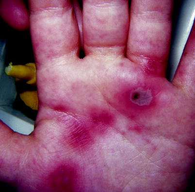
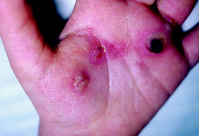
The lesions of primary cutaneous aspergillosis begin as tender erythematous or purpuric macules or papules that progress to violaceous edematous plaques, often with hemorrhagic bullae. Lesions tend to be large (centimeters) and single or a few grouped lesions if multiple. Lesions then develop dark centers and maintain a peripheral brightly purpuric rim. They may then ulcerate or form black necrotic eschars. The bullous phase of invasive aspergillosis is important to recognize, as potassium hydroxide (KOH) preparation of the hemorrhagic blister roof can provide an immediate presumptive diagnosis.
Less common presentations or morphologies of primary cutaneous aspergillosis include otomycosis
Primary cutaneous aspergillosis in HIV-positive patients is most commonly due to A. fumigatus .
Skin manifestations of secondary or disseminated aspergillosis are uncommon and occur in less than 10% of patients with invasive aspergillosis. However, cutaneous lesions may be the presenting sign of disseminated disease.
Skin biopsy specimens from Aspergillus infected patients demonstrate acute angle, regularly septate, dichotomously branching hyphal elements that may be angioinvasive. The histopathologic differential diagnosis includes several other opportunistic fungi including Fusarium , Scopulariopsis , Pseudallescheria , and Penicillium which appear identical to Aspergillus in tissue. In primary cutaneous disease, the inflammatory infiltrate is superficial or superficial and deep. In secondary cutaneous aspergillosis, the infiltrate is in the deep dermis and subcutaneous fat; intravascular thrombosis with masses of hyphae may be seen. While a skin biopsy is needed to prove invasive disease, isolation in culture of the organism from tissue specimens is needed for definitive identification of the fungus.
Enzyme linked immunoassay (ELISA) testing for the galactomannan antigen may aid in the diagnosis of invasive aspergillosis. Galactomannan is a carbohydrate component of the Aspergillus cell wall that is released during hyphal growth. It requires angioinvasion of the organism to be detected and can be tested in serum, brocheoalveolar lavage fluid, or cerebral spinal fluid. The sensitivity of the test ranges from 65% to 90% and the specificity is 90%; thus, a negative result rules out disease while a positive result requires a second positive to confirm true infection. False negatives occur if patients are tested while on antifungal agents or if there is limited angioinvasion. False positives occur with loss of integrity of gastrointestinal mucosa (mucositis, neonates, GVHD, cytotoxic chemotherapy) or with the use of piperacillin-tazobactam. It is not as widely used as the galactomannan ELISA.
Primary cutaneous aspergillosis has a more favorable prognosis than secondary or disseminated disease. Surgical excision in addition to systemic antifungal therapy may be beneficial for primary cutaneous aspergillosis. Systemic treatment is with antifungal therapy such as amphotericin B, itraconazole, voriconazole, or an echinocandin. Fluconazole is not an effective treatment for aspergillosis.
Figure 1.1.
Purpuric papules and a gray flat bullae due to Aspergillus flavus on the palm of a 7-year-old with acute lymphocytic leukemia (ALL) on the 15th day of chemotherapy, febrile with an absolute neutrophil count of 60 on triple antibiotics
Figure 1.2.
Forty-eight hours later, the papules became pustular and the bullae developed a gray-black necrotic base

