• Complain

Antonio Cardesa Pieter J. Slootweg Nina Gale - Pathology of the Head and Neck

Here you can read online Antonio Cardesa Pieter J. Slootweg Nina Gale - Pathology of the Head and Neck full text of the book (entire story) in english for free. Download pdf and epub, get meaning, cover and reviews about this ebook. City: Berlin;Heidelberg;New York, year: 1988, publisher: Springer;Churchill Livingstone, genre: Home and family. Description of the work, (preface) as well as reviews are available. Best literature library LitArk.com created for fans of good reading and offers a wide selection of genres:

Romance novel Science fiction Adventure Detective Science History Home and family Prose Art Politics Computer Non-fiction Religion Business Children Humor

Choose a favorite category and find really read worthwhile books. Enjoy immersion in the world of imagination, feel the emotions of the characters or learn something new for yourself, make an fascinating discovery.

Antonio Cardesa Pieter J. Slootweg Nina Gale Pathology of the Head and Neck

Pathology of the Head and Neck: summary, description and annotation

We offer to read an annotation, description, summary or preface (depends on what the author of the book "Pathology of the Head and Neck" wrote himself). If you haven't found the necessary information about the book — write in the comments, we will try to find it.

Antonio Cardesa Pieter J. Slootweg Nina Gale: author's other books


Who wrote Pathology of the Head and Neck? Find out the surname, the name of the author of the book and a list of all author's works by series.

Pathology of the Head and Neck — read online for free the complete book (whole text) full work

Below is the text of the book, divided by pages. System saving the place of the last page read, allows you to conveniently read the book "Pathology of the Head and Neck" online for free, without having to search again every time where you left off. Put a bookmark, and you can go to the page where you finished reading at any time.

Light

Font size:

Reset

Interval:

Bookmark:

Make
Springer-Verlag Berlin Heidelberg 2016
Antonio Cardesa , Pieter J. Slootweg , Nina Gale and Alessandro Franchi (eds.) Pathology of the Head and Neck 10.1007/978-3-662-49672-5_1
1. Benign and Potentially Malignant Lesions of the Squamous Epithelium and Squamous Cell Carcinoma
Nina Gale 1
(1)
Faculty of Medicine, Institute of Pathology, University of Ljubljana, Korytkova 2, 1000 Ljubljana, Slovenia
(2)
Department of Anatomic Pathology, Hospital Clinic, University of Barcelona, Villarroel 170, 8036 Barcelona, Spain
Nina Gale (Corresponding author)
Email:
Nina Zidar
Email:
Antonio Cardesa
Email:
Alfons Nadal
Email:
1.1
1.2
1.2.1
1.2.2
1.3
1.4
1.4.1
1.4.2
1.5
1.5.1
1.5.2
1.5.3
1.5.4
1.5.5
1.5.6
1.5.7
1.5.8
1.6
1.7
1.7.1
1.7.2
1.7.3
1.7.4
1.7.5
1.7.6
1.7.7
1.8
1.8.1
1.8.2
1.8.3
1.8.4
1.8.5
1.8.6
1.8.7
1.8.8
Keywords
Oral cavity Larynx Squamous cell papilloma and related lesions Squamous intraepithelial lesions Modified Ljubljana classification Squamous cell carcinoma and subtypes Second primary tumours Spread and metastasising Molecular carcinogenesis
1.1 Introduction
The chapter is focused on the three main groups of lesions of the covering squamous epithelium of the oral cavity and larynx. The first part treats squamous cell papillomas and related, viral-induced lesions with the main stress on laryngeal recurrent papillomatosis.
The second part is dedicated to squamous intraepithelial lesions (SILs), which still represent one of the most controversial topics in oral and laryngeal pathology. The modified Ljubljana classification provides clear morphological criteria for defining the prognostic groups of SILs squamous intraepithelial lesions and could acts as a model to a unified classification of the head and neck region. The third and the most extensive part is devoted to invasive squamous cell carcinoma (SCC) and its nine variants, including spindle cell carcinoma, verrucous carcinoma, papillary SCC, basaloid squamous cell carcinoma, non-keratinizing human papillomavirus positive SCC, adenoid squamous cell carcinoma, adenosquamous carcinoma and lymphoepithelial carcinoma. The second primary SCCs, which have a much lower 5-year survival than the primary tumors in the head and neck region, are also discussed. All manners of spreading and metastasising of the SCCs are widely discussed with the point on significant predictors of patients survival. The chapter concludes with a comprehensive review of the already known molecular events in carcinogenesis of head and neck SCC.
1.2 Squamous Cell Papilloma and Related Lesions
General considerations
Benign, exophytic, papillary or verrucous lesions of the squamous epithelium of the oral cavity, oropharynx and larynx include similar entities, such as squamous cell papilloma (SCP), verruca vulgaris (VV), condyloma acuminatum (CA) and focal epithelial hyperplasia (FEH), also known as Hecks disease. However, not every papillary lesion in these areas can be reliably placed into one of the listed categories. It seems that the majority of lesions are similar variants of papillary proliferations, all induced by infections with different genotypes of human papillomaviruses (HPV), showing more or less overlapping clinical and morphological attributes but different biological behaviour, ranging from rather inconspicuous to potentially life threatening. Classification of these changes into infectious (VV, CA, FEH) and neoplastic (SCP) is thought to be fairly inconsistent and not well founded. Papillary lesions, except laryngeal papillomatosis, generally have a favourable outcome.
1.2.1 Oral Squamous Cell Papilloma, Verruca Vulgaris, Condyloma Acuminatum and Focal Epithelial Hyperplasia
Definition
SCP, the most frequent papillary lesion of the oral cavity and oropharynx, is characterised as an exophytic papillary lesion, composed of fibrovascular projections covered by a benign proliferation of the squamous epithelium and induced by HPV infection.
VV is a rare intraoral lesion resembling its dermal counterpart, characterised as a solitary or multiple papules with verrucous surface and histologically classified as a wart-like hyperplasia of the squamous epithelium.
CA are usually larger than SCP, multiple, dome-shaped nodular lesions, resembling anogenital CA, which mainly appear on the lips and soft palate.
FEH is a rare oral lesion of children characterised by multiple sessile or elevated papules, usually distributed over the buccal, labial and tongue mucosa.
Epidemiology
SCP is most frequently located on the tongue and soft palate but may appear on any epithelial surface of the oral cavity [].
VV rarely occurs in the oral cavity; frequently affected sites are the labial mucosa of the lower lip and the vermilion border of both lips. The lesions, which are seen mainly in children, result from autoinoculation of HPV from VV on the fingers [].
CA is a rare, sexually transmitted lesion of adults. Common locations of CAs are the lips, tongue and gingival.
FEH is a rare, HPV-induced, contagious disease, initially described among the Native American population. FEH have also recently been published from other parts of the world. Small multiple lesions occur on the labial and buccal mucosa and tongue. The disease commonly occurs in children and young adults [].
Etiology and pathogenesis
Oral mucosa can be contaminated by HPV by various pathways, including sexual contacts, autoinfection and perinatal infection. Low-risk HPVs, which mainly induce the whole spectrum of oral papillary lesions, are also present in healthy persons; the prevalence of HPV detection in normal oral mucosa ranges from 0.6 to 81 % [].
Macroscopy
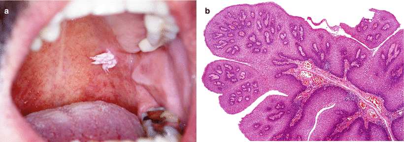
SCP is usually a single, pedunculated, white or pink lesion, consisting of fingerlike projections of the oral mucosa (Fig. ].
Fig 11 Oral squamous cell papilloma a Whitish papillary lesion of the - photo 1
Fig. 1.1
Oral squamous cell papilloma. ( a ) Whitish papillary lesion of the palate (Courtesy of Dr. J. Fischinger, Ljubljana, Slovenia). ( b ) Projections of fibrovascular stroma are covered by parakeratotic squamous epithelium
VVs are frequently multiple, rough-surfaced sessile lesions of whitish colour.
CAs are characterised as small, sessile pink papules, which can combine into a larger cauliflower lesion.
FEHs are sessile, well-demarcated, round or ovoid flat lesions; they can appear in clusters and measure up to 10 mm in diameter;
Microscopy
SCP is composed of narrow papillary projections of soft fibrous stroma covered by keratotic or parakeratotic, hyperplastic squamous epithelium, usually with normal maturation. Rarely, basalparabasal cell hyperplasia is seen, as well as an increased number of mitoses (Fig. ).
Koilocytosis, the only visible cytopathic effect of HPV infection, caused by viral replication in the upper intermediate and superficial zone of the squamous epithelium, is rarely visible in SCPs. The characteristics of koilocytes are described in the paragraph on laryngeal papillomatosis. VV shows similar histological features, but peripheral papillary projections are usually centrally inverted and koilocytosis and the granular layer are prominent. The base of the lesion is usually broad and flat. CA is histologically described as a broad papillary proliferation with koilocytosis and parakeratosis on the surface of the epithelium and bulbous rete ridges with a possible extension into the ducts of the minor salivary glands [).
Next page
Light

Font size:

Reset

Interval:

Bookmark:

Make

Similar books «Pathology of the Head and Neck»

Look at similar books to Pathology of the Head and Neck. We have selected literature similar in name and meaning in the hope of providing readers with more options to find new, interesting, not yet read works.


Reviews about «Pathology of the Head and Neck»

Discussion, reviews of the book Pathology of the Head and Neck and just readers' own opinions. Leave your comments, write what you think about the work, its meaning or the main characters. Specify what exactly you liked and what you didn't like, and why you think so.