Springer International Publishing Switzerland 2016
E. Edmund Kim , Hyung-jun Im , Dong Soo Lee and Keon Wook Kang Atlas and Anatomy of PET/MRI, PET/CT and SPECT/CT 10.1007/978-3-319-28652-5_1
1. Atlas and Anatomy of PET/MR
E. Edmund Kim 1, Hyung-Jun Im 2, Dong Soo Lee 3 and Keon Wook Kang 4
(1)
Department of Radiological Sciences, School of Medicine University of California at Irvine, Irvine, CA, USA
(2)
Department of Nuclear Medicine, Seoul National University, Seoul, Republic of Korea
(3)
Department of Nuclear Medicine and Department of Molecular Medicine and Biopharmaceutical Sciences, Seoul National University, Seoul, Republic of Korea
(4)
Department of Nuclear Medicine and Cancer Research Institute, Seoul National University, Seoul, Republic of Korea
Keywords
PET/MR 18F-Fludeoxyglucose (FDG) Oncology Anatomy Cancer
After the huge success of hybrid positron emission tomography/computed tomography (PET/CT), there has been a continuous effort to develop a hybrid positron emission tomography/magnetic resonance image (PET/MR) machine. Recently, a magnetic fieldcompatible PET component has been developed by a substituting photomultiplier tube (PMT) for an avalanche photodiode (APD) or silicon multiplier (SiPM). This enables development and commercialization of PET/MR. Commercial simultaneous PET/MR is now seeking clinical validation. A simultaneous PET/MR system has several intrinsic advantages over a PET/CT system, including a lower radiation dose, higher soft tissue resolution of anatomic images, and the possibility of using a novel multifunctional PET/MR probe. In addition, there is the potential for the simultaneous acquisition of an anatomic image and PET. PET/MR has a higher soft tissue resolution than PET/CT; therefore the image reader should be well trained in reading normal anatomy and abnormal findings in MR for the proper reading of PET/MR. There are many MR books and atlases available to help understand and read MR images; however, there are few PET/MR atlases. This chapter includes typical PET/MR cases of patients with malignant tumors in the area of the brain, head and neck, chest, abdomen, pelvis, and musculoskeletal system. In each case, pathologic findings and essential surrounding normal structures for interpretation are indicated and named [].
1.1 Brain
1.1.1 Case 1
A male patient, age 75, presented with worsening dizziness and weakness in both legs for 1 month. A tumorous condition in the brain was suspected on brain CT and therefore 18F-Fludeoxyglucose (FDG) PET/MR was used.
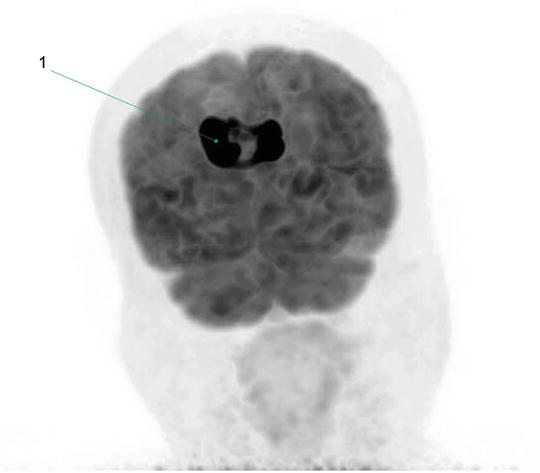
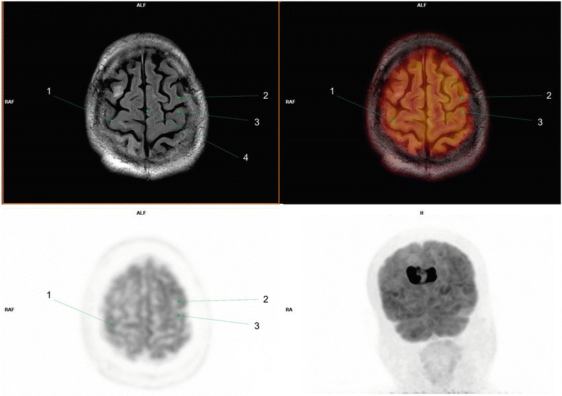
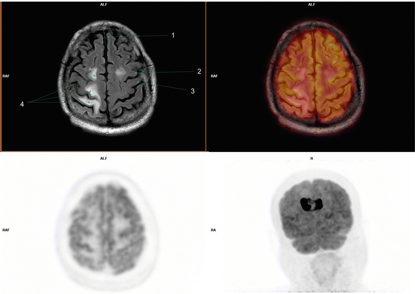
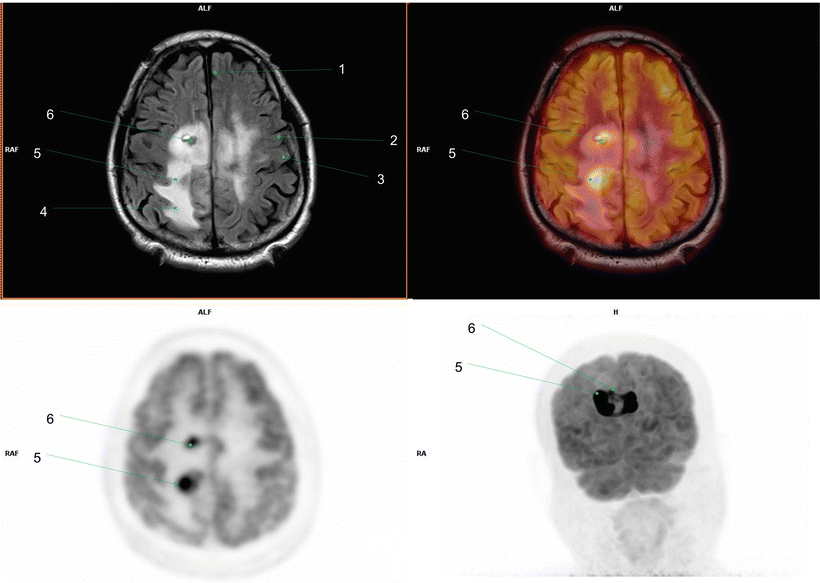
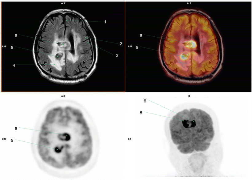
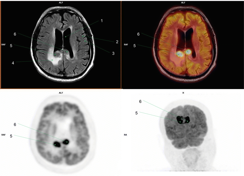
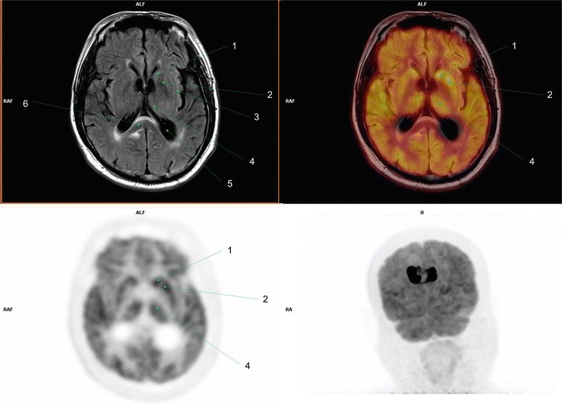
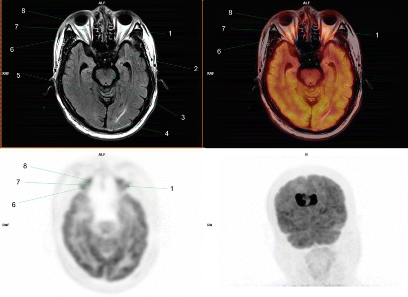
Brain FDG PET/MR revealed a well-enhanced mass with intense metabolic activity involving the body of the corpus callosum. There was no abnormal lesion with increased metabolic activity in the rest of the imaged body. Primary central nervous system (CNS) lymphoma was suspected, and stereotaxic biopsy revealed a diffuse large B-cell lymphoma [).
Fig. 1.1
(1) Primary central nervous system lymphoma
Fig. 1.2
(1) Right postcentral gyrus(2) Left precentral gyrus(3) Left postcentral gyrus(4) Falx cerebri
Fig. 1.3
(1) Left superior frontal gyrus(2) Left precentral gyrus(3) Left postcentral gyrus(4) Peritumoral edema
Fig. 1.4
(1) Left superior frontal gyrus(2) Left precentral gyrus(3) Left postcentral gyrus(4) Peritumoral edema(5) Primary central nervous system lymphoma in right parietal white matter(6) Primary central nervous system lymphoma in right frontal white matter
Fig. 1.5
(1) Left superior f rontal gyrus(2) Left precentral gyrus(3) Left postcentral gyrus(4) Peritumoral edema(5) Pri mary central nervous system lymphoma involving right parietal white matter(6) Primary central nervous system lymphoma involving corpus callosum
Fig. 1.6
(1) Left latera l ventricle(2) Left precentral gyrus(3) Left postcentral gyrus(4) Peritumoral edema(5) Primary central nervous system lymphoma involving posterior corpus callosum(6) Primary central nervous system lymphoma involving posterior corpus callosum
Fig. 1.7
(1) Left cauda te nucleus(2) Left putamen(3) Left insular cortex(4) Left thalamus(5) Left lateral ventricle trigone(6) Corpus callosum (splenium)
Fig. 1.8
(1) Left lat eral rectus muscle(2) Left temporal cortex(3) Midbrain(4) Occipital cortex(5) Cerebellar vermis(6) Right lateral rectus muscle(7) Optic nerve(8) Right medial rectus muscle
1.1.2 Case 2
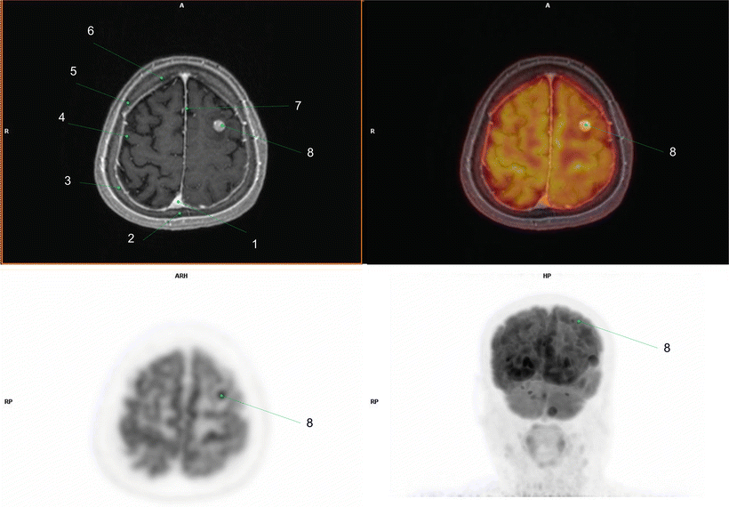
A 74-year-old female patient suffering from a tingling sensation in her right hand and aphasia for 10 days was examined. A tumorous condition in the brain was suspected on brain CT, and FDG PET/MR was performed.
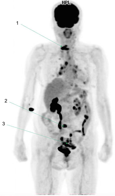
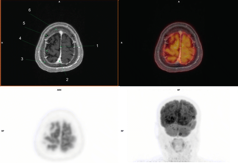
Brain FDG PET/MR revealed multiple round-shaped enhancing hypermetabolic nodules with peritumoral edema in the left frontal parietal lobes and cerebellum that were appeared to be metastases. In whole-body FDG PET/MR, hypermetabolic bone spinal lesions were found in C7 and L5, suggesting bone metastases. Also, an infiltrative lesion with increased metabolic activity was found in the sigmoid colon. Subsequent colonoscopy and colonoscopic biopsy revealed sigmoid colon cancer. The metastatic lesions were considered to have originated from the colon cancer [).
Fig. 1.9
(1) Bone metas tasis in cervical spine(2) Bone metastasis in lumbar spine(3) Sigmoid colon cancer
Fig. 1.10
(1) Falx cerebri(2) Superior sagittal sinus(3) Right parietal bone(4) Right postcentral gyrus(5) Right coronal suture(6) Frontal bone
Fig. 1.11
(1) Superior s agittal sinus(2) Sagittal suture(3) Right parietal bone(4) Right postcentral gyrus(5) Right coronal suture(6) Frontal bone(7) Falx cerebri(8) Metastasis in left frontal lobe

