Fast Facts: Early Breast Cancer

| Jayant S VaidyaMBBS MS DNB FRCS PhD FRCS(Gen) Professor of Surgery and Oncology
Consultant Surgeon
Whittington, Royal Free and UCL Hospitals
Scientific Director, Clinical Trials Group
Division of Surgery and Interventional Science
University College London, UK |

| Vivek PatkarMBBS MS MRCS Fellow of European Board of Breast Surgery
Senior Research Fellow
University College London, UK |
Declaration of Independence
This book is as balanced and as practical as we can make it.
Ideas for improvement are always welcome:

Fast Facts: Early Breast Cancer
First published January 2017
Text 2017 Jayant S Vaidya and Vivek Patkar
2017 in this edition Health Press Limited
Health Press Limited, Elizabeth House, Queen Street, Abingdon,
Oxford OX14 3LN, UK
Tel: +44 (0)1235 523233
Book orders can be placed by telephone or via the website.
To order via the website, please go to: fastfacts.com
For telephone orders, please call +44 (0)1752 202301.
Fast Facts is a trademark of Health Press Limited.
All rights reserved. No part of this publication may be reproduced, stored in a retrieval system, or transmitted in any form or by any means, electronic, mechanical, photocopying, recording or otherwise, without the express permission of the publisher.
The rights of Jayant S Vaidya and Vivek Patkar to be identified as the authors of this work have been asserted in accordance with the Copyright, Designs & Patents Act 1988 Sections 77 and 78.
The publisher and the authors have made every effort to ensure the accuracy of this book, but cannot accept responsibility for any errors or omissions.
For all drugs, please consult the product labeling approved in your country for prescribing information.
Registered names, trademarks, etc. used in this book, even when not marked as such, are not to be considered unprotected by law.
A CIP catalogue record for this title is available from the British Library.
ISBN 978-1-910797-12-9
eISBN 978-1-910797-24-2
Vaidya JS (Jayant)
Fast Facts Breast Cancer/
Jayant S Vaidya, Vivek Patkar
The authors wish to thank Professor David Joseph, Dr Alison Jones, Professor Michael Baum and Professor Harvey Schipper for their contributions to previous editions of Fast Facts: Breast Cancer from which some of this text is adapted: Vaidya JS, Joseph D. Fast Facts: Breast Cancer, 5th edn. Oxford: Health Press Limited, 2014.
Medical illustrations by Dee McLean, London, and
Anna-Maria Dutto, Withernsea, UK.
Printed in the UK with Xpedient Print.
In developed countries, breast cancer is usually diagnosed at an early stage when it is still confined to the breast and regional nodes. Here, we focus on stage 0 (ductal carcinoma in situ), I, II and IIIA disease, with a concise overview that will aid understanding of the risk of developing breast cancer, the essentials of diagnosis and preoperative assessment and the current approach to treatment and follow-up.
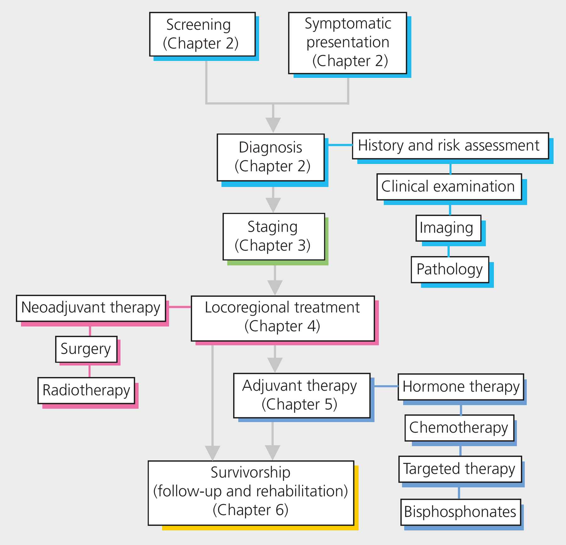
Use Fast Facts: Early Breast Cancer to follow clear diagnostic and treatment pathways, from screening and symptomatic presentation, through triple assessment diagnosis, to treatment and follow-up.
Definitions
Early breast cancer refers to cancer that has not spread beyond the breast or the axillary lymph nodes. This includes ductal carcinoma in situ (DCIS; stage 0) and stage I, IIA, IIB and IIIA invasive breast cancers (see ). The term invasive is unique to breast cancer: in no other cancer is such tautology used. It simply means that cancer cells have crossed the basement membrane of the duct and can potentially spread. It is only used because of the popularity of the terms ductal or lobular carcinoma in situ (DCIS and LCIS) in which cancer cells have not crossed the basement membrane; such lesions, if pure, are theoretically not a risk to life. DCIS and LCIS are most commonly identified by mammographic screening or as a chance finding after a biopsy of a benign lesion. Ductal intraepithelial neoplasia (DIN) would be the preferred name and would remove the unnecessary taboo of cancer when these lesions are diagnosed.
There are no histopathological or molecular markers to predict the progression from DCIS to invasive disease with certainty. However, the risk of invasive cancer is believed to be increased by nine- to elevenfold in a woman in whom DCIS is treated by removal of the affected area alone.
Incidence and mortality
Breast cancer is the most common form of cancer among women, with an estimated 1.67 million new cases diagnosed worldwide in 2012.
Structure of the breast and surrounding tissues
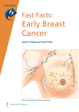
The breast is composed of glandular and adipose tissue in varying proportions. The glandular tissue consists of 1520 lobes containing numerous lobules, linked by ductules (). The ductules combine to form the lactiferous ducts, which open into the lactiferous sinuses and empty through the nipple. The breast is enclosed in two layers of fibrous tissue connected by Coopers ligaments, which give it its characteristic shape. Invasion of Coopers ligaments by cancer and cicatrization shortens the ligaments, leading to the classic early sign of dimpling of skin when the pectoralis is contracted or when the patient has their arms raised when bent forward.
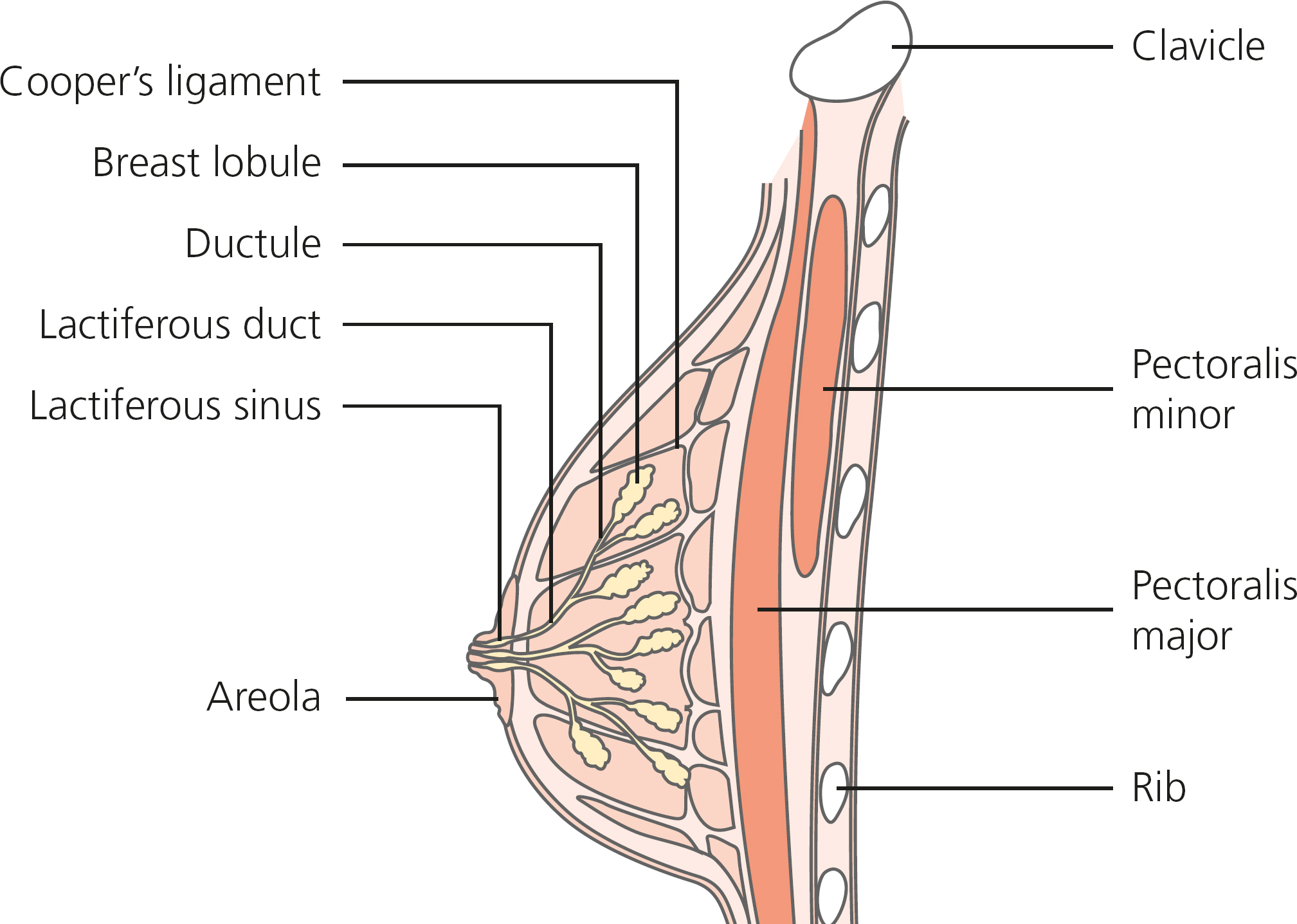
Figure 1.1 Structure of the normal breast.
The lymphatics of the breast tissue converge in the subareolar plexus of Sappy, and then drain into the axilla (armpit).
Pathophysiology
Most breast cancers are epithelial tumors, arising from either the milk-producing glands (lobular carcinomas) or, more commonly, from the draining ducts (ductal carcinomas); only a small number are non-epithelial involving the stroma or soft tissues.
Ductal carcinomas account for over 90% of breast cancers. Lobular carcinomas account for approximately 8% of breast cancers. Such tumors may occur at several sites, either in the same breast or in both breasts. They can be hard to diagnose, as their diffuse nature and relative radiolucency mean that they often do not show up on mammograms.
Phyllodes tumors are relatively rare stromal tumors that only very rarely exhibit the malignant features of a true sarcoma. Clinically and on imaging they resemble fibroadenomas, although they are often larger.
Receptor status. Estrogen and progesterone are important regulators of normal breast growth and development and play important roles in the pathogenesis of breast cancer. The hormone receptors in some breast cancers promote DNA replication and cell division when estrogen or progesterone bind to them (e.g. an estrogen-receptor positive [ER+] tumor), while the presence of the receptor for human epidermal growth factor 2 (HER2) correlates with a poorer prognosis at any given stage of cancer. These receptors provide valuable therapeutic targets, as blocking them can stop cancer growth and may even lead to complete regression.
Next page
