John W. Krakauer - Broken Movement: The Neurobiology of Motor Recovery after Stroke
Here you can read online John W. Krakauer - Broken Movement: The Neurobiology of Motor Recovery after Stroke full text of the book (entire story) in english for free. Download pdf and epub, get meaning, cover and reviews about this ebook. year: 2021, publisher: The MIT Press, genre: Home and family. Description of the work, (preface) as well as reviews are available. Best literature library LitArk.com created for fans of good reading and offers a wide selection of genres:
Romance novel
Science fiction
Adventure
Detective
Science
History
Home and family
Prose
Art
Politics
Computer
Non-fiction
Religion
Business
Children
Humor
Choose a favorite category and find really read worthwhile books. Enjoy immersion in the world of imagination, feel the emotions of the characters or learn something new for yourself, make an fascinating discovery.

- Book:Broken Movement: The Neurobiology of Motor Recovery after Stroke
- Author:
- Publisher:The MIT Press
- Genre:
- Year:2021
- Rating:4 / 5
- Favourites:Add to favourites
- Your mark:
- 80
- 1
- 2
- 3
- 4
- 5
Broken Movement: The Neurobiology of Motor Recovery after Stroke: summary, description and annotation
We offer to read an annotation, description, summary or preface (depends on what the author of the book "Broken Movement: The Neurobiology of Motor Recovery after Stroke" wrote himself). If you haven't found the necessary information about the book — write in the comments, we will try to find it.
Broken Movement: The Neurobiology of Motor Recovery after Stroke — read online for free the complete book (whole text) full work
Below is the text of the book, divided by pages. System saving the place of the last page read, allows you to conveniently read the book "Broken Movement: The Neurobiology of Motor Recovery after Stroke" online for free, without having to search again every time where you left off. Put a bookmark, and you can go to the page where you finished reading at any time.
Font size:
Interval:
Bookmark:
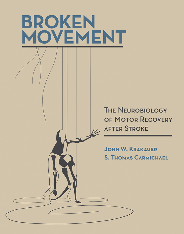
Broken Movement
The Neurobiology of Motor Recovery after Stroke
John W. Krakauer and S. Thomas Carmichael
The MIT Press
Cambridge, Massachusetts
London, England
2017 Massachusetts Institute of Technology
All rights reserved. No part of this book may be reproduced in any form by any electronic or mechanical means (including photocopying, recording, or information storage and retrieval) without permission in writing from the publisher.
This book was set in Times New Roman by Westchester Publishing Services. Printed and bound in the United States of America.
Library of Congress Cataloging-in-Publication Data
Names: Krakauer, John W., author. | Carmichael, S. Thomas.
Title: Broken movement : the neurobiology of motor recovery after stroke / John W. Krakauer and S. Thomas Carmichael.
Description: Cambridge, MA : The MIT Press, [2017] | Includes bibliographical references and index.
Identifiers: LCCN 2017021092 | ISBN 9780262037228 (hardcover : alk. paper)
Subjects: LCSH: Cerebrovascular diseasePatientsRehabilitation. | Motor learning.
Classification: LCC RC388.5 .K67 2017 | DDC 616.8/1dc23
LC record available at https://lccn.loc.gov/2017021092
I dedicate this book to my husband, Omar, who patiently weathered my nocturnal writing binges, mountains of papers and books, and demands to be fed.
JWK
I dedicate this book to my wife, Diana, whose unfailing calm and understanding balances impatience, impertinence, and indelicacy.
TC
- ICF levels and measures of arm paresis. Stroke measures mapped onto the International Classification of Functioning, Disability, and Health (ICF) framework. Different measures capture stroke-related motor disability at different levels; for example, quantifiable measures of movement kinematics are more specific at the level of motor impairment while self-reported measures assess the impact of stroke at higher levels of disability. Clinical measures are not specific and span disability levels with varying degrees of overlap.
- Task-related activity for a pyramidal tract neuron (PTN) producing postspike facilitation (PSF) in the first dorsal interosseous (FDI) muscle. (A) PTN discharges for sixteen repetitions of the precision grip task showing an increase in activity before and during the light and heavy force conditions. Rectified and summed electromyography (EMG), recorded at the same time (shown at right). (B) Corresponding task-related analysis for the same PTN and muscle during the power grip. Adapted from Muir and Lemon (1983).
- Redundancy in arm joint coordination for the same kinematic goal. The goal here is to draw a circle on the wall using a laser pointer. This can be accomplished by rotating about the wrist, elbow, or shoulder. An almost infinite number of combinations are possible.
- Decrease in work area of the arm as a function of various levels of limb support. Work areas in the left paretic limb are inverted to facilitate the comparison with the right nonparetic limb. Data from a single patient; axes units are in meters. Adapted from Sukal et al. (2007).
- Trajectory and joint kinematics during two kinds of reach. Top Pane: Schematic of (A) reaching condition, target position 1 for reach out and target position 2 for reach up and (B) shoulder individuation task (elbow and wrist individuation tasks not shown). Left Pane: Overlaid single trials for index finger paths and associated excursions of the shoulder, elbow, and wrist joints from both reaching conditions (up and out). (A) Control index finger paths for reaches out and up. (B) Control joint angle plots during the reach out. (C) Control joint angle plots during the reach up. (D) Hemiparetic subject index finger paths. (E) Hemiparetic subject joint angle plots during the reach out. (F) Hemiparetic subject joint angle plots during the reach up. Solid black circles: target. Asterisk: index end-point position or time of index end point prior to corrective submovements. Angular excursions are graphed as follows: bold dashed traces, shoulder angular excursion; narrow solid traces, elbow angular excursion; narrow dashed traces, wrist angular excursion. Right Pane: Shoulder, elbow, and wrist individuation movements graphed over time from start of movement to maximum angular excursion in the instructed joint. For a healthy control (left) and a hemiparetic subject (right). Bold dashed traces, shoulder angular excursion; narrow solid traces, elbow angular excursion; narrow dashed traces, wrist angular excursion. Adapted from Zackowski et al. (2004).
- Isometric finger individuation task. Mean deviation from baseline in the passive fingers plotted against the force generated by the active finger. The higher the slope of the relationship, the worse the individuation ability. Data from seven patients and seven healthy controls. Adapted from Xu et al. (2017).
- Upregulation of the ipsilateral reticulospinal tract after stroke. Responses of identified motor neurons to ipsilateral pyramidal tract stimulation. (A) Schematic showing the lesioned pyramidal tract (PT, dashed line) and potential pathways descending through the ipsilateral (intact) PT that might contribute new connections. (B) Quantitative measurements of input to motor neurons from the ipsilateral PT. Data are presented separately according to the muscle group that the motor neuron innervated: forearm flexors, forearm extensors, and intrinsic hand muscles. Light-shaded bars relate to control and dark to lesioned animals. Bars are grouped depending on if the responses are monosynaptic or disynaptic. The product of amplitude and incidence measures connection strength. There were no significant differences between control and lesioned animals. (C) Schematic showing the lesioned pyramidal tract (PT, dashed line) and potential pathways descending through the ipsilateral (intact) reticulospinal tract (medial longitudinal fasciculus [MLF]) that might contribute new connections. (D) Same quantitative measurements of input to motor neurons from the reticulospinal tract. There were significant differences between control and lesioned animals for forearm flexors and intrinsic hand muscles for both monosynaptic and disynaptic responses Adapted from Zaaimi et al. (2012).
- Comparison of hand paths in the left and right arms of controls and patients with mild hemiparesis (all subjects for both groups were right-handed). Left hemiparesis is associated with end-point errors, whereas right hemiparesis is associated with directional trajectory errors. LHC: Left Healthy Control; RHC: Right Healthy Control; LHD: Left Hemisphere Damage; RHD: Right Hemisphere Damage. Adapted from Mani et al. (2013).
- Differential impact of posture-dependent hypertonia on the control of limb trajectory and end-posture stabilization after stroke. (A) Topography of posture-dependent flexor forces acting on the hand of one stroke survivor instructed to relax as a planar robot held the hand at various sampled locations. Each stroke survivor yielded a unique posture map. Also shown are target locations for cued movements from the home position into regions of HIGH (dark gray areas) and LOW (light gray areas) postural bias forces. (B) Schematic view of an out-and-back reversal movement produced by a stroke survivor reaching without concurrent visual feedback into a HIGH bias force region of workspace. Gray dots: trajectory locations of hand speed maxima during the outward and return phases. Shading: reversal area. (C) Average reversal areas for neurally intact (NI) control subjects, less-impaired stroke survivors (Fugl-Meyer Motor Assessment [FMA] > 30) and more impaired survivors (FMA < 30). Reversal areas were normalized by the square of the target movement distance to account for individualized differences in active range of motion and consequent differences in target distance. (D) Overhead view of an out-and-hold reach hand path produced by a stroke survivor without concurrent visual feedback. Inset: corresponding hand speed profile during stabilization against small, 3N peak-to-peak robotic force perturbations applied after hand speed dropped below 20 percent peak speed. The ability to stabilize the hand at the target was quantified using the sum of measured hand displacements for the one-second period after the onset of perturbations. Solid line: portion of the trajectory used to compute terminal hand displacement for this movement. (E) Average terminal hand displacement for the same NI control subjects, less-impaired stroke survivors (FMA > 30), and more impaired survivors (FMA < 30). Error bars: 1 SEM. Adapted from Simo et al. (2013).
Font size:
Interval:
Bookmark:
Similar books «Broken Movement: The Neurobiology of Motor Recovery after Stroke»
Look at similar books to Broken Movement: The Neurobiology of Motor Recovery after Stroke. We have selected literature similar in name and meaning in the hope of providing readers with more options to find new, interesting, not yet read works.
Discussion, reviews of the book Broken Movement: The Neurobiology of Motor Recovery after Stroke and just readers' own opinions. Leave your comments, write what you think about the work, its meaning or the main characters. Specify what exactly you liked and what you didn't like, and why you think so.

