Lifes Blueprint
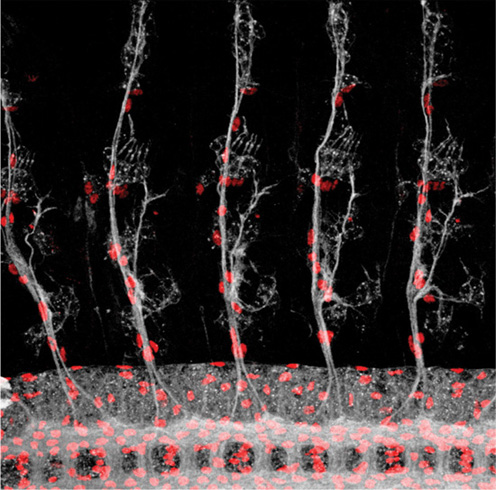
Trees of embryonic neurons
Lifes Blueprint
The Science and Art of Embryo Creation
Benny Shilo

Published with assistance from the Alfred P. Sloan Foundation Public Understanding of Science and Technology Program and from the foundation established in memory of Amasa Stone Mather of the Class of 1907, Yale College.
Copyright 2014 by Benny Shilo. All rights reserved. This book may not be reproduced, in whole or in part, including illustrations, in any form (beyond that copying permitted by Sections 107 and 108 of the US Copyright Law and except by reviewers for the public press), without written permission from the publishers.
Yale University Press books may be purchased in quantity for educational, business, or promotional use. For information, please e-mail (UK office).
Printed in China.
Library of Congress Cataloging-in-Publication Data
Shilo, Benny, 1951 author.
Lifes blueprint : the science and art of embryo creation / Benny Shilo.
pp ; cm.
Includes bibliographical references and index.
ISBN 978-0-300-19663-4 (clothbound : alk. paper)
I. Alfred P. Sloan Foundation. Public Understanding of Science and Technology Program, sponsoring body. II. Title.
[DNLM: 1. Embryonic Development. 2. Embryonic Structurescytology. QS 604]
QM601
612.64dc23 2014014622
A catalogue record for this book is available from the British Library.
This paper meets the requirements of aNSI/NISo Z39.481992 (Permanence of Paper).
10 9 8 7 6 5 4 3 2 1
Frontispiece: Trees of embryonic neurons. The sensory neurons of the peripheral nervous system of the fruit fly embryo connect to the central nervous system, seen as a ladder at the bottom. They transmit information from external sense organs and internal muscle stretch receptors. The structure of the neurons is repeated accurately at every segment of the embryo. Some of the neural cell nuclei are marked in red, and nerve axons in gray (M. Silies and C. Klmbt, Mnster University)
To my parents,
Prof. Moshe Shilo and Dr. Mira Shilo,
who embedded in me the passion for science
and introduced me to the wonders of biology
Contents
Preface
During fertilization, the genetic contents of the egg and sperm, each carrying half of the genetic material of the mother and father, respectively, are united in a single cell. This unicellular embryo will give rise to a complete multicellular organism, with its enormous complexity of tissues and multiple types of cells that will appear different from one another and carry out diverse and distinct roles. All the information for this elaborate process is carried within the DNA of that initial single cell that was formed on fertilization, and all cells of the embryo will contain exactly the same genetic information ().
The long-standing human desire to understand the instructions and rules of forming an embryo and to unravel lifes blueprint stems from many sources. First and foremost, we want to understand how we as humans, and all other organisms, build our complex bodies. How do genes shape animals? What are the instructions? How are they carried out in such a reliable and reproducible fashion? Can we use this knowledge to better understand pathological situations in which the genetic program goes awry? And, thinking to the future, can we use the information gained in order to instruct cells to generate tissues and organs that could be used as spare parts?
As we set out to explore these fundamental questions, more specific and operational issues come to mind. Are cells preprogrammed by the genome to generate distinct cell types, or do cells have a choice of assuming different fates? How do they decide which fate to choose from the arsenal of tissues they are capable of forming? Does every species have its own rules and logic of embryonic development, or, alternatively, can we identify common themes among disparate species? The scientific discoveries of the past three decades have revolutionized our knowledge and understanding of embryonic development in all multicellular organisms. This book conveys these striking findings and the emerging rules of executing development.
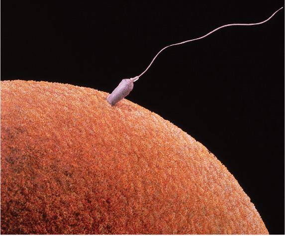
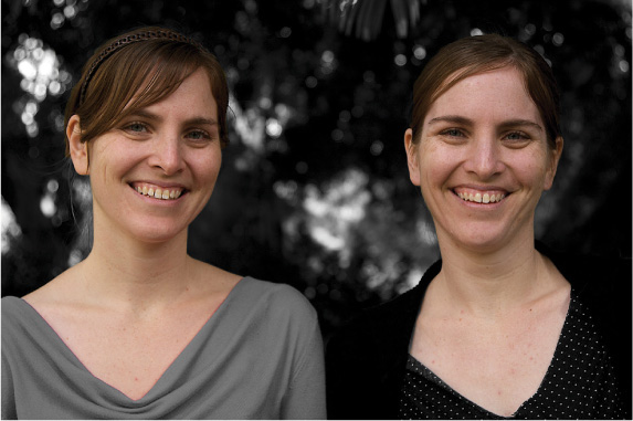
Figure 1. All cells in our body carry identical genetic information
Upon fertilization, the nuclei of the sperm and egg join, and the first cell of the embryo is formed. Throughout the numerous divisions that will follow, all descendants will contain the same genetic legacy. This information dictates the shape of the embryo, as indicated by the fact that identical twins carrying the same genetic material are so similar.
Top: Scanning electron micrograph of fertilized human egg (F. Leroy/Science Source); bottom: Identical twins (B. Shilo, Rehovot, Israel)
The analysis of embryonic development relies strongly on microscopic images that we obtain and analyze. These images include fluorescent multicolor depictions of different embryonic tissues, scanning electron microscope images, and movies of live tissues and embryos showing dynamic processes in real time. The images are not only highly informative but often also aesthetically striking. Their visual beauty is often central in attracting many researchers to the field.
For me, these visual scientific stimulations are combined with another personal passion. Since my high school years, I have been interested in photography and how we view and depict our surroundings. In my early work, I felt more comfortable photographing still objects and how they interact with light. In the past fifteen years, as a result of frequent trips to foreign destinations, including the countries of the Far East and India, and possibly also as a consequence of a more mature view of the world, I have concentrated on photographing people. I try to portray universal aspects that unite people of all races and ages, as well as the unique features of individuals in their surroundings.
To visually present the emerging concepts of embryonic development in this book, I have assembled scientific images of diverse organisms that display the central concepts of embryogenesis from the laboratories of colleagues and from my own lab. Each of these images is paired with an image from our macro world taken by me, highlighting a similar theme and reflecting my personal metaphors. In seeing these juxtaposed images, I hope that readers will be able to understand each paradigm in an intuitive way, letting our everyday experiences and recollections resonate with and inform our comprehension of the scientific image.
It is interesting to note that the inception of the word cell as a biological term was formulated by Robert Hooke in 1665 and was based on a visual analogy to the macro world. Hooke looked at the bark of a cork oak using a microscope he developed. He saw the regular shapes of the nonliving cell walls and noted that they resemble the cells in which monks live. I envisage that through the pictorial analogies, the reader will feel like a cell within the developing embryo ().
Next page

