Acknowledgments
All chapters in Breathe Easy have been revised multiple times after review and comments by several individuals. I am grateful for the insights provided by Susan McNeely and Carl Small based on their perspectives and personal experiences. Special thanks to Alex H. Gifford, MD, assistant professor of medicine at Geisel School of Medicine at Dartmouth; Scott Astle, RRT, and clinical coordinator of cardiorespiratory services at Valley Regional Hospital in Claremont, New Hampshire; Mary Anne Riley, RRT, and program coordinator of pulmonary rehabilitation programs at Springfield Hospital in Springfield, Vermont, and at Cheshire Medical Center in Keene, New Hampshire; and Drew Carter, senior sales representativerespiratory for Lincare Inc., in Enfield, New Hampshire.
I appreciate the creative efforts of Kyle Morrison (
( 1 )
How to Breathe
Breathing in I calm my body. Breathing out I smile. Dwelling in the present moment, I know this is a wonderful moment!
Thich Nhat Hanh, Vietnamese Buddhist monk and author (1926 )
Breathing is essential to life. One purpose of each and every breath is to take in oxygen. However, it is equally important that we get rid of carbon dioxidea waste product of our bodyduring exhalation. To understand how to breathe, it is helpful to review the parts (anatomy) and workings (function) of the respiratory system. This information provides a framework for knowing about various lung conditions if you, or a loved one, has a breathing problem.
How to breathe depends on the interactions between the brain and the respiratory system. These areas are connected by nerves (called the nervous system) that enable communication by electrical signals that travel back and forth. Sensors in the respiratory system provide information to the brain about breathing, and the brain controls how fast and how deep to breathe.
This chapter also reviews specific techniques for how the brain can ease breathing discomfort and distress. These include mindful breathing and breathing retraining. Pursed-lips breathing is a useful strategy that provides some relief as well as a sense of control when breathlessness develops. Lastly, yoga includes breathing as an integral part to control the mind and the body.
Anatomy of the Respiratory System
The respiratory system starts in your nose and mouth and ends in the air sacs (alveoli; ).
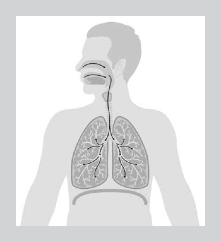
Figure 1.1 Pathway for inhaled air to enter the nose or mouth and pass through the windpipe (trachea) and the breathing tubes (airways) to reach the air sacs (alveoli). (Based on iStock.com images from elenabs and kowalska-art)
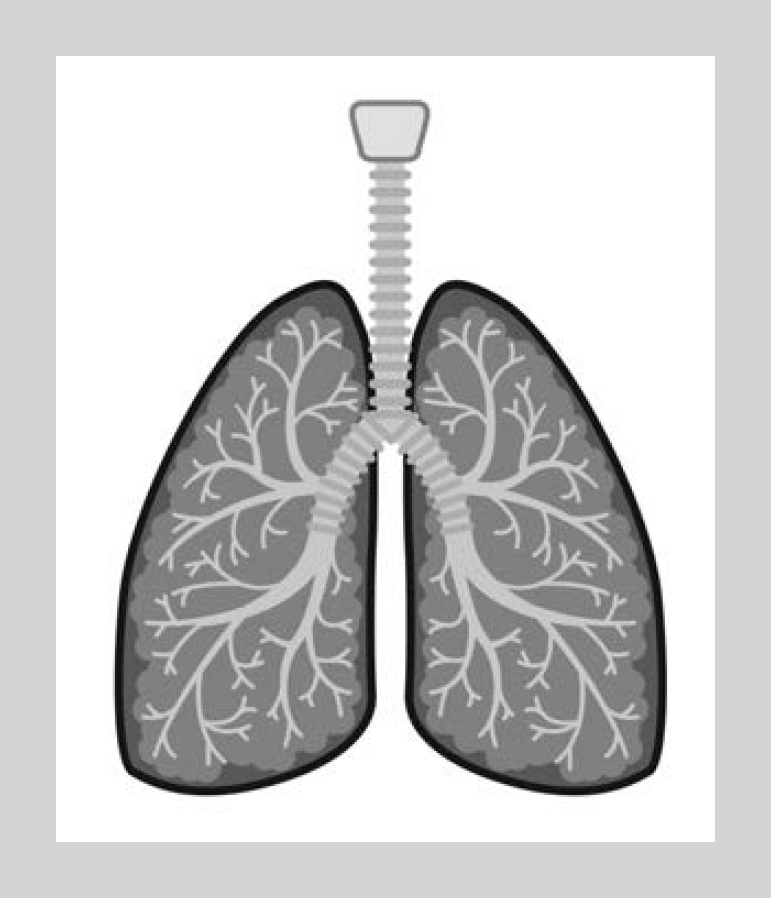
Figure 1.2 The vocal cords, windpipe (trachea), and breathing tubes (airways), which divide twenty-three times to end in air sacs (alveoli). (iStock.com/elenabs)
Table 1.1 The upper respiratory system
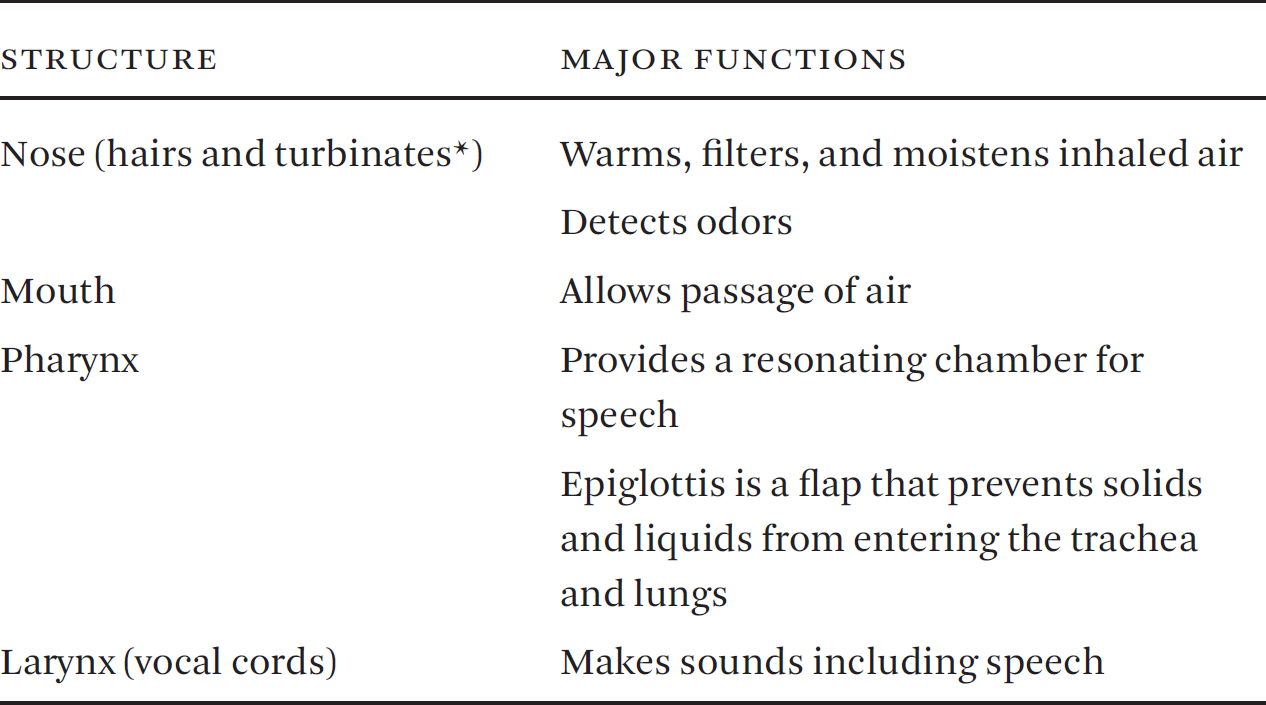
*Turbinates are long, narrowed, and curled bone shelves shaped like a seashell that protrudes into the breathing passage of the nose. Turbinates provide humidity for the lining of the nose that is needed for smell.
Table 1.2 The lower respiratory system

The respiratory system is divided into upper and lower parts. The structures and major functions of the upper respiratory system are described in .
The main structures and their functions of the lower respiratory tract are listed in .
Normal Breathing
Most people do not think about their breathing as it is an unconscious action. Although you breathe ten to twelve times each minute, this happens without even a thought or concern. The size of each breath, called tidal volume, is about five hundred milliliters for an average size adult. This equals two cups of air (). Think about it: you breathe in and out two cups of air each breath that you take!
How does this happen? A group of nerve cells are located at the base of the brain that regularly sends electrical signals through nerves to the breathing muscles ().
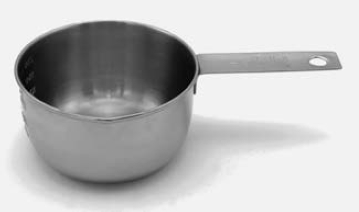
Figure 1.3 One-cup measuring container. The average breath is two cups of air. (iStock.com/sarahdoow)
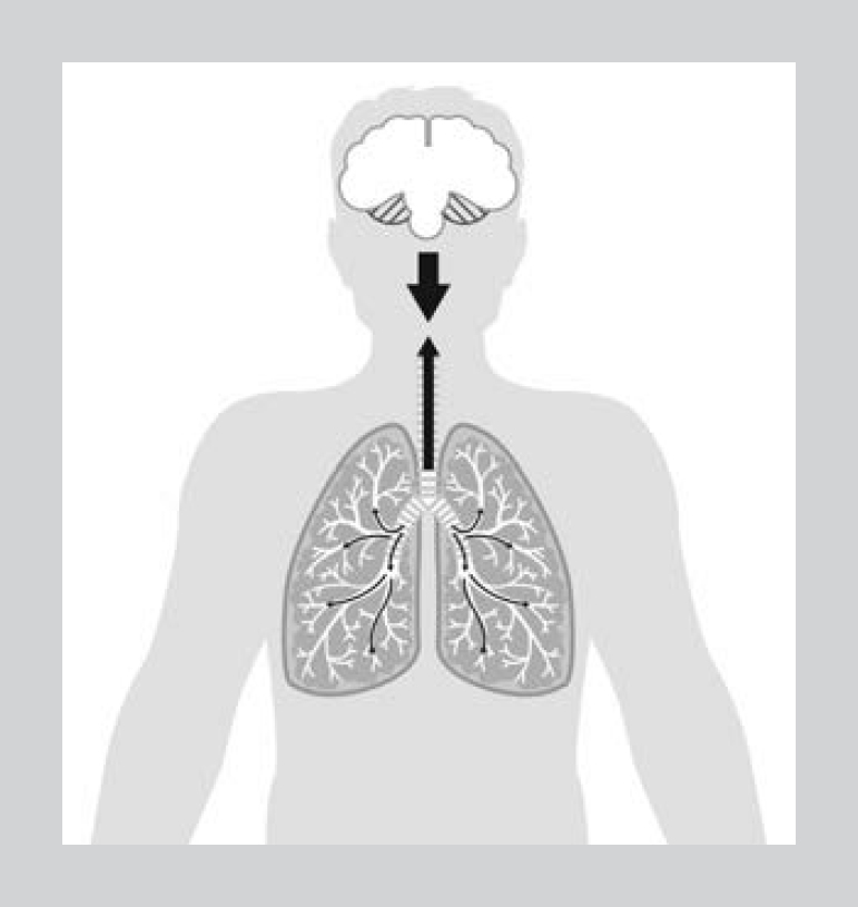
Figure 1.4 A group of nerves located in the lower part of the brain (brain stem) sends signals automatically to the breathing muscles that control how often we breathe and how deep. The upper part of the brain (cerebral cortex) has the ability to voluntarily control breathing. (Based on iStock.com images from elenabs and kowalska-art)
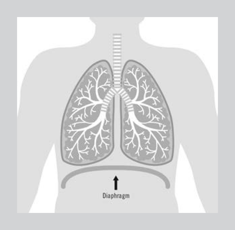
Figure 1.5 Shows the diaphragm, which is shaped like a dome and separates the chest and stomach (abdomen) cavities. (iStock.com/elenabs)
This is called the respiratory center and functions as a pacemaker for breathing. The right and left phrenic nerves (phrenic means mind in Latin) start in the neck (cervical nerves three through five) and pass down through the chest to each side of the diaphragm. The diaphragm is a muscle that is shaped like a dome. It is the main breathing muscle and separates the chest and the stomach (abdomen; ).
The electrical signals that travel through the phrenic nerves tell the diaphragm when and how much to shorten (contract). When this happens, the dome-shaped diaphragm moves down toward the stomach cavity like a piston. Electrical signals also travel through other nerves to muscles located between ribs (called intercostal muscles). When the intercostal muscles shorten (contract), the ribs are lifted out to assist the diaphragm to breathe in air ().
If there is an even greater need to breathe in more air, electrical signals are also sent to neck muscles (called scalene and sternocleidomastoid muscles). When this happens the neck muscles shorten (contract) and lift the upper chest to breathe in more air ().
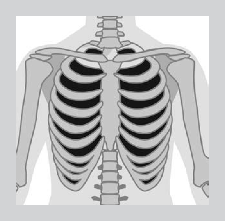
Figure 1.6 Each intercostal muscle connects an upper and lower rib on both sides of the chest. When these muscles shorten (contact), the ribs are lifted to help breathe in air. (iStock.com/elenabs)
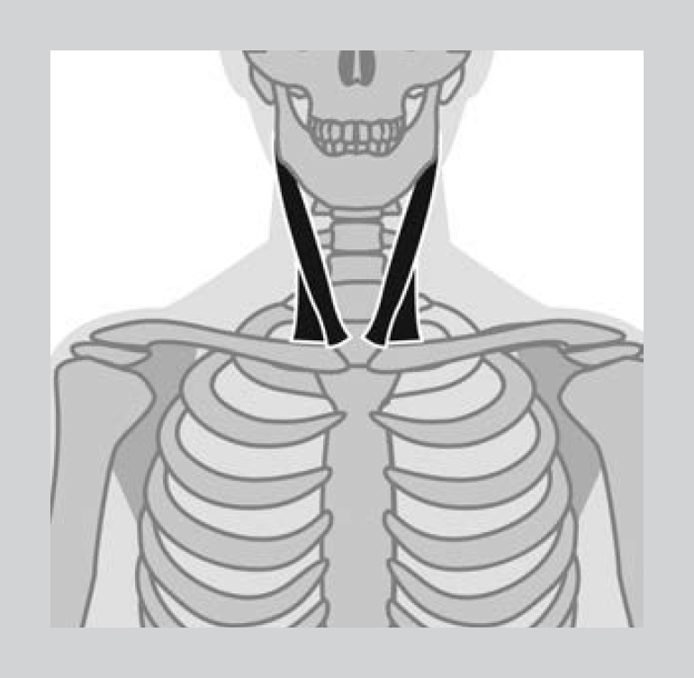
Figure 1.7 Diagram of sternocleidomastoid muscles that connect the head and the upper chest. These muscles can assist breathing if necessary. (iStock.com/elenabs)
Voluntary Control of Breathing
The brain is able to override automatic breathing. This is called voluntary control as shown in . For example, you can decide to blow up a balloon or to hold your breath. This ability to voluntarily breathe more or breathe less when you want is unique to the respiratory system. No other part of the body can do this. Although the heart has a group of nerve cells that function as a pacemaker in order to control how many times the heart beats each minute, the brain cannot make the heart beat faster or slower!
Next page
