Musculoskeletal MRI
Third Edition
Nancy M. Major, MD
Professor of Radiology and Orthopedics, Division of Musculoskeletal Imaging, University of Colorado School of Medicine, Aurora, Colorado
Mark W. Anderson, MD
Professor of Radiology and Orthopaedic Surgery, Harrison Distinguished Teaching Professor of Radiology, University of Virginia, Charlottesville, Virginia
Clyde A. Helms, MD
Durham, North Carolina
Phoebe A. Kaplan, MD
Niwot, Colorado
Robert Dussault, MD
Niwot, Colorado

Copyright
Elsevier
1600 John F. Kennedy Blvd.
Ste 1800
Philadelphia, PA 19103-2899
MUSCULOSKELETAL MRI, THIRD EDITION ISBN: 978-0-323-415606
Copyright 2020 by Elsevier, Inc. All rights reserved.
No part of this publication may be reproduced or transmitted in any form or by any means, electronic or mechanical, including photocopying, recording, or any information storage and retrieval system, without permission in writing from the publisher. Details on how to seek permission, further information about the Publishers permissions policies and our arrangements with organizations such as the Copyright Clearance Center and the Copyright Licensing Agency, can be found at our website: www.elsevier.com/permissions.
This book and the individual contributions contained in it are protected under copyright by the Publisher (other than as may be noted herein).
Notice
Practitioners and researchers must always rely on their own experience and knowledge in evaluating and using any information, methods, compounds or experiments described herein. Because of rapid advances in the medical sciences, in particular, independent verification of diagnoses and drug dosages should be made. To the fullest extent of the law, no responsibility is assumed by Elsevier, authors, editors or contributors for any injury and/or damage to persons or property as a matter of products liability, negligence or otherwise, or from any use or operation of any methods, products, instructions, or ideas contained in the material herein.
Previous editions copyrighted 2009, and 2001.
Library of Congress Control Number: 2019944072

Publisher: Russell Gabbedy/Joslyn T. Chaiprasert-Paguio
Senior Content Development Manager: Ellen M. Wurm-Cutter
Publishing Services Manager: Deepthi Unni
Project Manager: Beula Christopher
Design Direction: Ryan Cook
Printed in China
Last digit is the print number: 9 8 7 6 5 4 3 2 1
Dedication
To Austin, your support and humor saw me through this book. To my mother and father, I am forever grateful for your love and support. To my dear friend, and co-author, Mark, your partnership and patience during this third edition was invaluable. And to Kenneth, thank you is not enough. Forever and always.
Nancy M. Major
To the residents and fellows who have made me a better radiologist and my job worth doing. To Nancy whose tireless efforts have made this a much-improved edition. And to Amy whose unwavering love and infinite patience have made me a better person.
Mark W. Anderson
Preface
Nancy M. Major, MD ; Mark W. Anderson, MD
Since the first edition of Musculoskeletal MRI was published in 2001, much has changed in the realm of musculoskeletal MRI. Scores of new articles have been published on every anatomic area, and new imaging techniques have been developed. Although we have attempted to incorporate these advances in the current edition, we stand firm in our original maxim that less is more when it comes to a text explaining the basics. Therefore we resolved to not let the size of this work increase dramatically. Almost every chapter has been significantly updated. Many new figures have been added and many of the original figures have been replaced by better examples using more current techniques. The text has been updated to reflect current research and practices.
Working on the third edition of Musculoskeletal MRI made us a little better at what we do, which is to practice and teach musculoskeletal MRI, and we are certain it also will help improve the skills of every reader of this book.
Basic Principles of Musculoskeletal MRI
Chapter Outline
- What Makes a Good Image?
- Lack of Motion
- Signal and Resolution
- Tissue Contrast
- Pulse Sequences
- Fat Saturation
- Gadolinium
- MR Arthrography
- Musculoskeletal Tissues
- Bone
- Normal Appearance
- Most Useful Sequences
- Pitfalls
- Articular Cartilage
- Normal Appearance
- Most Useful Sequences
- Fibrocartilage
- Normal Appearance
- Useful Sequences: Meniscus
- Pitfalls
- Useful Sequences: Glenoid or Acetabular Labrum
- Tendons and Ligaments
- Normal Appearance
- Most Useful Sequences
- Pitfalls
- Muscle
- Normal Appearance
- Useful Sequences
- Synovium
- Normal Appearance
- Useful Sequences
- Pitfalls
- Applications
- Suggested Reading
Although a detailed understanding of nuclear physics is not necessary to interpret magnetic resonance imaging (MRI) studies, it also is unacceptable to read passively whatever images you are given without concern for how the images are acquired or how they might be improved. Radiologists should have a solid understanding of the basic principles involved in acquiring excellent images. This chapter describes the various components that go into producing high-quality images, stressing the fundamental principles shared by all MRI scanners.
Every machine is different. Clinical scanners are now available at strengths ranging from 0.2 tesla (T) to 3.0T. Additionally, each vendor has its own language for describing its hardware, software, and scanning parameters, and an entire chapter could be devoted to deciphering the terms used by different manufacturers. Time spent learning the details of your machine with your technologists or physicists would be time well spent. If you are interested, read one of the excellent discussions of MRI physics in articles or other textbooks because, for the most part, in this book we leave the physics to the physicists.
What Makes a Good Image?
Lack of Motion
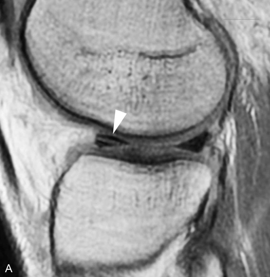
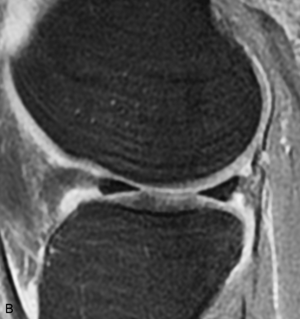
Motion is one of the greatest enemies of MRI (). It can arise from a variety of sources, such as cardiac motion, bowel peristalsis, and respiratory movement. For most musculoskeletal applications, motion usually stems from body movement related to patient discomfort. Patient comfort is of paramount importance because even if all the other imaging parameters are optimized, any movement would ruin the entire image.


