Page List
Photographing the Unseen World

Photographing
the Unseen World
Art and Techniques
Adrian Davies

First published in 2020 by
The Crowood Press Ltd
Ramsbury, Marlborough
Wiltshire SN8 2HR
www.crowood.com
This e-book first published in 2020
Adrian Davies 2020
All rights reserved. This e-book is copyright material and must not be copied, reproduced, transferred, distributed, leased, licensed or publicly performed or used in any way except as specifically permitted in writing by the publishers, as allowed under the terms and conditions under which it was purchased or as strictly permitted by applicable copyright law. Any unauthorised distribution or use of this text may be a direct infringement of the authors and publishers rights, and those responsible may be liable in law accordingly.
British Library Cataloguing-in-Publication Data
A catalogue record for this book is available from the British Library.
ISBN 978 1 78500 704 0
Acknowledgements
A book like this, covering a wide range of subject matter would not have been possible without the help of numerous friends and colleagues, for help, advice and occasional modelling! Andrea Blum, Bjorn Rorslett, and various members of the ultravioletphotography.com forum; Stephen Bleksley; Paul Buff Inc.; Matt Cass; Lucie Cash, Royal Mail; Paul Colley; Dr Jonathan Crowther; Hannah Davies (and Jack); Janice Davies; Adam Davies, Maidenhead Aquatics; Robin Davies; Bryony Davies; Miles Herbert, Captive Light Photography; Kat Jack; Yealand Kalfayan; Nicola Musgrave BSc (Hons) MSc., Eurofins Forensic Services; Phred Petersen, RMIT University; Paul Reynolds, Sigma Imaging (UK); Martin Sanders; Hugh Turvey Hon FRPS, FRSA; Paul Wilson; Michael Robbs
Chapter 1
Introduction
F rom its earliest beginnings, photography has been used to help visualize the unseen events that are either too slow or too fast for the human eye to perceive, to record subjects that are visible only in areas of the spectrum outside the human range of sensitivity, or record detail that is too small for the human eye to see. There are, and have been, many imaging techniques for visualizing the invisible, including X-ray and gamma-ray imaging, various medical scanning techniques such as MRI and CAT scans, Kirlian photography, schlieren photography, electron and light microscopy, thermal imaging and various forms of satellite imaging, though most of these techniques are unavailable to most readers, being either too dangerous, too expensive or requiring unattainable pieces of equipment.
This book is concerned with those techniques that anyone with a good knowledge of photography can achieve with minimal outlay. It will look at ultraviolet (UV) and infrared (IR) photography, high speed and time-lapse photography, close-up, macro and photography with the aid of a microscope, and photography using polarized light and other related techniques. Some of the applications will utilize two or more of the techniques in combination, and do not fit into neat categories.
The very act of viewing a subject through a magnifying glass can reveal previously unseen details within a subject, and the higher the power of the magnifying glass, or microscope, the more detail will be revealed. In particular, 3D electron microscopy not only reveals otherwise invisible detail, but also produces, at times, stunningly beautiful images. Electron microscopes are very expensive, and well outside the scope of this book, but a brief discussion of light microscopy will be given in .
The whole of life lies in the verb seeing.
Pierre Teilhard de Chardin
X-ray images of everyday subjects often reveal beautiful and startling details, such as the carnivorous pitcher plants shown here, alongside UV images of the same plant.
Schlieren photography is another technique used by scientists, and restricted to laboratory use, but capable of revealing heat and pressure patterns around suitable subjects such as the heat emitted from the soldering iron shown here. The cover image for John Martyns Solid Air album, from 1973, was a schlieren image of the heat emitted from a hand.
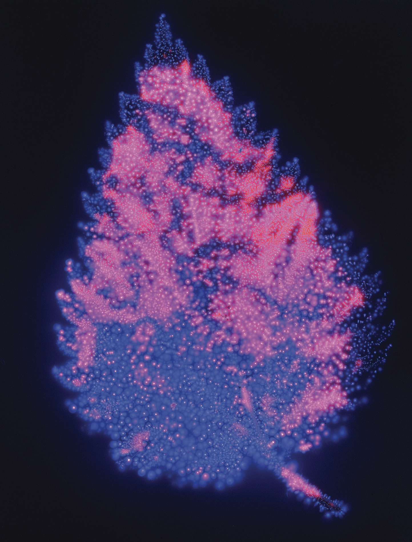
Fig. 1.1
Kirlian image of stinging nettle (Urtica dioica) leaf. The leaf was placed on a sheet of 5 4 transparency film in a dark room, which itself was placed on a metal sheet. A high voltage electrical current was passed through the metal sheet, resulting in a Kirlian image.
Home made Kirlian imaging device. 5" 4" Fujichrome film. Exposure approx. 30 seconds.
Kirlian photography (see ) is another visualization technique, used to capture the phenomenon of electrical coronal discharges (often referred to as the aura around a subject). It is named after Semyon Kirlian, who, in 1939, accidentally discovered that if an object is placed on a photographic plate that is connected to a high-voltage source, it produces an image. It became popular in the 1980s, and various claims for the technique were made with regard to it being able to predict illness or other issues, or reveal the natural aura around a subject. There is little evidence for these claims, and the technique has largely fallen out of use.
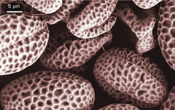
Fig. 1.2
Image of lilac (Syringa sp.) pollen at a magnification of 3,000, using a scanning electron microscope.
Authors: Yapryntsev, A.D; Baranchikov, A.E; Ivanov, V.K. This file is licensed under the Creative Commons Attribution 4.0 International license.

Fig. 1.3
Several different imaging techniques were used to illustrate different aspects of a carnivorous plant (Nepenthes spp.), resulting in very different images and differing information about the subject in .Conventional image of Nepenthes spathulata (copelandii truncata) shot with visible light.
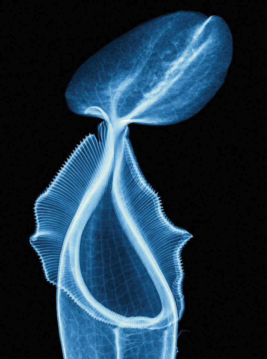
Fig. 1.4
X-ray image of carnivorous pitcher plant (Nepenthes ventricosa talangensis) yielding a truly beautiful image, showing remarkable clarity and detail. Unfortunately, the equipment required is not available to most people.
Image copyright Hugh Turvey, Hon FRPS, FRSA.
UV photography, in particular, is nowadays helping us get an idea of how other animals see the world. As early as 1864, Hermann Vogel, a German chemist, photographed a woman using a photographic plate that at that time was only sensitive to UV and blue light, using a simple, uncoated lens that transmitted good quantities of UV. The processed image showed several black spots on the womans face, invisible in normal daylight. A few days later the woman was diagnosed with smallpox. The significance of this UV reflectance image was not realized for another forty years or so.

