Thorsten Johnson , Christian Fink , Stefan O. Schnberg and Maximilian F. Reiser (eds.) Medical Radiology Dual Energy CT in Clinical Practice 10.1007/174_2010_43 Springer-Verlag Berlin Heidelberg 2011
Physical Background
Thorsten R. C. Johnson 1 and Willi A. Kalender 2
(1)
Department of Clinical Radiology, University of Munich, Grosshadern Hospital, Marchioninistrasse 15, 81377 Munich, Germany
(2)
Institute of Medical Physics, University of Erlangen, Henkestrasse 91, 91052 Erlangen, Germany
Thorsten R. C. Johnson
Email:
Abstract
There had been attempts to utilize spectral information for tissue characterization soon after the invention of Computed Tomography, but only recently Dual Energy CT has achieved a significant role in clinical radiology.
To perform Dual Energy CT, it is necessary to generate x-rays with different energies, mostly as polychromatic spectra. On the other hand, the detector has to be capable to differentiate x-ray quanta of different energies. There are four technical approaches to meet these requirements, of which the Dual Source CT, the rapid voltage switching and the layer detector technology are available or being implemented.
To obtain relevant diagnostic information, there have to be substances with spectral properties which reflect the pathology by their presence or distribution. Most important is the photoelectric effect of elements like uric acid, iron, calcium, iodine or xenon gas, which are present in pathological structures or can be administered as contrast material. The identification and quantification of these elements can be used to diagnose several diseases.
History
First attempts to use spectral information in computed tomography date back to the late 1970s (Millner et al. ). Today, there are several technical approaches to dual-energy CT.
X-ray Spectra
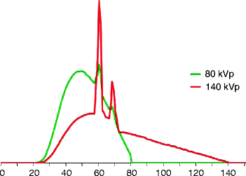
Generally, there are three requirements for spectral CT imaging: First, X-ray sources providing X-ray quanta with different energies are necessary. Obviously, this can be achieved optimally when using two separate X-ray sources emitting different photon energies. However, this criterion is basically also met by single X-ray sources, because they generally provide polychromatic spectra. The X-ray spectrum of a normal tube with a tungsten anode consists of a continuous part, the Bremsstrahlung, and discrete peaks according to the energy levels of the electrons in the shell of the tungsten atoms. Figure shows the X-ray spectra that are obtained from an X-ray tube operated at 140 and 80 kV. Obviously, the area under the curve for equivalent tube currents differs by a factor of about 45, and the tube current needs to be adapted to obtain a similar output of quanta from both tubes. The higher energy spectrum is dominated by the characteristic lines of the tungsten anode, while the lower energy spectrum mainly consists of Bremsstrahlung. In this case, the mean photon energies are 71 and 53 keV, respectively, for the primary spectra. It is desirable to have as little overlap as possible between the spectra; therefore, the lowest and highest potentials offered by the respective CT scanner are always used for dual-energy acquisitions. A tube voltage lower than 80 kV is not useful because too much of the quanta would be absorbed by the human body; values higher than 140 kV are generally not available and would result in very low soft tissue contrast that likely would contribute very little to tissue differentiation. Obviously, it is not possible to obtain monochromatic X-rays with todays tube technology; monochromatic X-ray sources so far do not provide a sufficient output of quanta for clinical applications. An exception is monochromatic synchrotron radiation that can be used for experimental setups. As synchrotrons are far too large for use in a rotating gantry, the object has to be rotated between the source and the detector, i.e., it is not possible to use them for clinical imaging.
Fig. 1
Spectra of the Straton tube at 140 and 80 kV potential. The peaks represent the characteristic lines of the tungsten anode and the continuous spectrum is a result of Bremsstrahlung. The mean photon energies are 53 and 71 keV, respectively
Detector Technology
The second requirement is that the detector has to be able to differentiate quanta of different energies. With technologies in use in clinical CT today, this is not directly possible with a single detector. The detector integrates the fluorescent light intensities of all photons detected during a single readout interval, but does not give account of their energy. Current approaches either rely on entirely separate X-ray sources and corresponding separate detectors, on reading out the projection data at different time points, or on using a two-layer or sandwich detector with different spectral sensitivities. In the near future, cadmium-based materials such as CdZnTe may serve as semiconductors for photon-counting detectors that resolve the energy of each individual photon. However, this detector technology cannot yet cope with the high photon flux required for clinical CT.
These first two requirements define the spectra of the X-ray sources and the corresponding detectors, representing the sensitivity of the systems for photons, detectors of different energies. The more these spectra differ, the stronger the contrast-to-noise ratio of the resulting spectral information, as long as both sufficient transmission and attenuation can be achieved in the human body at these photon energies.
Tissue Properties
The third requirement is a sufficient difference in spectral properties of the materials under investigation. The attenuation that is quantified in CT to characterize different body tissues is caused by three physical processes. Compton scatter is the largest component of attenuation. It is related to the electron density and not to the protons in the atomic core that would allow a material differentiation (McCullough ). Therefore, only elements with a considerable difference in Z values will be distinguishable by their spectral properties. This difference can be characterized with the so-called dual-energy index (DEI):
where x 80 is the CT value in HU at 80 kV tube potential and x 140 the value of the respective voxel at 140 kV (Johnson et al. ).
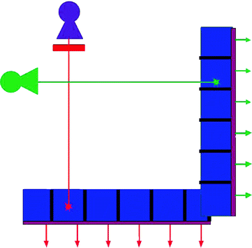
Fig. 2
Sketch of a dual-source CT system. Two tubes and detectors are mounted orthogonally. To obtain dual-energy datasets, the tubes are operated at different tube voltages, e.g., 80 and 140 kV ( green and violet ). Additionally, a filter (indicated in red ) can be applied to harden the high-energy spectrum
According to Alvarez and Macovski (), which is generally used in CT as a contrast agent and the distribution of which can be masked by the underlying tissue.
Dual-Source CT
At present, there are three different approaches to clinical dual-energy CT commercially available or under investigation. The most straightforward approach is dual-source CT with two X-ray sources running at different voltages with two corresponding detectors (Fig. ).