High-Performance Deformable Image Registration Algorithms for Manycore Processors
Copyright
Morgan Kaufmann is an imprint of Elsevier
225 Wyman Street, Waltham, MA, 02451, USA
First published 2013
Copyright 2013 Elsevier Inc. All rights reserved
No part of this publication may be reproduced or transmitted in any form or by any means, electronic or mechanical, including photocopying, recording, or any information storage and retrieval system, without permission in writing from the publisher. Details on how to seek permission, further information about the Publishers permissions policies and our arrangement with organizations such as the Copyright Clearance Center and the Copyright Licensing Agency, can be found at our website: www.elsevier.com/permissions
This book and the individual contributions contained in it are protected under copyright by the Publisher (other than as may be noted herein).
Notices
Knowledge and best practice in this field are constantly changing. As new research and experience broaden our understanding, changes in research methods, professional practices, or medical treatment may become necessary.
Practitioners and researchers must always rely on their own experience and knowledge in evaluating and using any information, methods, compounds, or experiments described herein. In using such information or methods they should be mindful of their own safety and the safety of others, including parties for whom they have a professional responsibility.
To the fullest extent of the law, neither the Publisher nor the authors, contributors, or editors, assume any liability for any injury and/or damage to persons or property as a matter of products liability, negligence or otherwise, or from any use or operation of any methods, products, instructions, or ideas contained in the material herein.
British Library Cataloguing-in-Publication Data
A catalogue record for this book is available from the British Library
Library of Congress Cataloging-in-Publication Data
A catalog record for this book is available from the Library of Congress
ISBN: 978-0-12-407741-6
For information on all MK publications visit our website at www.mkp.com

Biographies
James Shackleford is an assistant professor in the electrical and computer engineering department at Drexel University. Prior to joining Drexel, he was a postdoctoral researcher at Massachusetts General Hospital in the department of radiation oncology. Dr. Shackleford received his Ph.D. from Drexel University in 2011 for his work on GPU-accelerated medical image processing, implemented as part of the plastimatch project (www.plastimatch.org), a deformable registration toolkit for medical images maintained by Drs. Shackleford and Sharp. He has authored a chapter in NVIDIAs GPU Computing Gems (Emerald Edition) on the topic of accelerating deformable 3-D image registration using uniform cubic B-splines and this work has also been published as a featured article in the Physics in Medicine and Biology journal. His other research interests include nanoscale solid-state device physics.
Nagarajan Kandasamy is an associate professor in the electrical and computer engineering department at Drexel University, where he teaches and conducts research in the area of computer engineering, with specific interests in performance management, parallel computing, embedded systems, fault-tolerant computing, and computer architecture. He received his Ph.D. in 2003 from the University of Michigan. Prof. Kandasamy is a recipient of the National Science Foundation Early Faculty (CAREER) Award, as well as best paper awards at the 2006 and 2008 IEEE International Conferences on Autonomic Computing, for work focusing on the power and performance management of large-scale computer clusters.
Greg Sharp is a computer scientist and medical physicist at Massachusetts General Hospital. He received his Ph.D. in the department of electrical engineering and computer science from the University of Michigan in 2002, and currently holds an appointment of assistant professor of radiation oncology at Harvard Medical School. Prof. Sharps research interests include medical image computing, image-guided radiation therapy, and motion management.
Chapter 1
Introduction
Information in This Chapter:
Motivation for multicore CPU/GPU implementations
Applications of deformable registration
Algorithmic approaches to deformable registration
Organization of the book
1.1 Introduction
The fundamental step for combining three-dimensional (3D) geometric data is registration, which is the process of aligning two or more images that capture the geometric structure of the same scene, but in their own relative coordinate frames, into a common coordinate frame. The images themselves can be obtained at different times and from different viewpoints, using similar or different imaging modalities. Here, we focus on volumetric registration, where the images are pixel or voxel intensities arranged in a regular grid, and the relative alignment of multiple images must be found. Volumetric registration is often used in biomedical imaging, e.g., to track changes in a patients anatomy using images taken at different time points or to align stacks of microscopy data in either space or time.
A registration is called rigid if the motion or change is limited to global rotations and translations, and is called deformable if it includes complex local variations. One of the images is often called the static or reference image and the second image is the moving image, and registration involves spatially transforming the moving image to align with the reference image. When registering medical images, e.g., of a patients anatomy taken at different time points, one must account for deformation of the anatomy itself due to the patients breathing, anatomical changes, and so on.
Modern imaging techniques such as computed tomography (CT), positron emission tomography (PET), and magnetic resonance imaging (MRI) provide physicians with 3D image volumes of patient anatomy , thus allowing for precise tumor localization during the resection procedure.
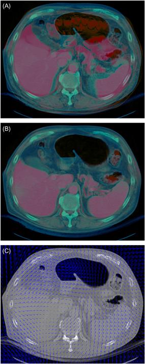
Figure 1.1 Computing organ motion via deformable registration. (A) A preoperative MRI image (in red) superimposed on an intraoperative CT image (in blue) before deformable registration. (B) The preoperative MRI superimposed on the intraoperative CT after deformable registration. (C) The deformation vector field (in blue) derived by the registration process superimposed on the intraoperative CT scan wherein the vector field quantitatively describes the organ motion between the CT and MRI scans.


