Rosalie E. Ferner , Susan M. Huson and D. Gareth R. Evans Neurofibromatoses in Clinical Practice 10.1007/978-0-85729-629-0_1 Springer-Verlag London Limited 2011
1. Neurofibromatosis 1
Rosalie E. Ferner 1, 2
(1)
Department of Neurology, Guys and St. Thomas NHS Foundation Trust, London, UK
(2)
Department of Clinical Neuroscience, Institute of Psychiatry, Kings College, London, UK
Abstract
Neurofibromatosis 1(NF1) is a common autosomal dominant condition that primarily involves the skin and the nervous system.
People with NF1 have an increased risk of developing benign and malignant tumors.
Neurofibromatosis 1
Neurofibromatosis 1(NF1) is a common autosomal dominant condition that primarily involves the skin and the nervous system.
People with NF1 have an increased risk of developing benign and malignant tumors.
History of Current Terminology
Descriptions of people allegedly suffering from neurofibromatosis date back to the first century AD. One of the more convincing reports comes from Tilesius (1793) who depicted a short man with a curved spine and a large head.
Table 1.1.
Diagnostic criteria for NF.
The clinical diagnosis is made when at least two of the following are present: |
A first degree relative with NF1 |
Six or more caf au lait patches >0.5 cm in children and >1.5 cm in adults |
Axillary or groin freckling |
Two or more neurofibromas of any type or one plexiform neurofibroma |
Two or more Lisch nodules (iris hamartomas) |
Optic pathway glioma |
Bony dysplasia of the sphenoid wing or |
Thinning of the long bone cortex with or without pseudarthrosis of the long bones |
Source: National Institutes of Health Consensus Development Conference Statement. Copyright 1988 American Medical Association. All rights reserved
Epidemiology
NF1 has a birth frequency of 1 in 2,5003,000 and a minimum prevalence of 1 in 45,000.
Mosaic NF1
This form of NF1 occurs in about 1 in 30,000 individuals and presents either as mild generalized disease that is clinically identical to NF1 or with segmental manifestations.).
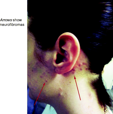
Figure 1.1.
Segmental NF1. Cutaneous neurofibromas around the left side of the face and neck in individual with no other signs of NF1.
Genetics of NF1
In 1990 the NF1 gene was identified on chromosome 17q11.2 and the protein product neurofibromin is found in high levels in the nervous system. People with NF1 are likely to develop benign and malignant tumors because neurofibromin loses its tumor suppressor function through gene mutation.
Genetics of Mosaic NF1
The NF1 mutation arises in the egg or sperm before fertilization in classical generalized NF1 and after fertilization in mosaic NF1.
Skin Manifestations of NF1
Skin problems are frequently the presenting manifestation of the disease and caf au lait patches, freckling, and neurofibromas form part of the diagnostic criteria of NF1.
Caf au lait Patches
Caf au lait patches usually develop at birth or in early infancy and are observed in 99% of people with NF1 by age of 3 years (Fig.
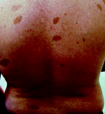
Figure 1.2.
Multiple caf au lait patches.
Freckling
Freckling is observed as hyperpigmented macules 13 mm in diameter in 85% of children after the age of 3 years.
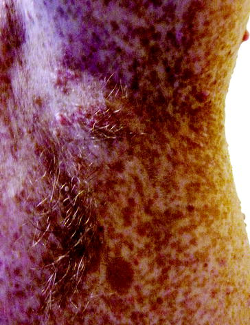
Figure 1.3.
Axillary freckling.
Campbell de Morgan Spots
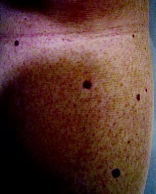
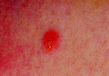
Campbell de Morgan spots are cherry red angiomas 13 mm in diameter and occur predominantly on the trunk and thighs (Fig. They are common in NF1 patients and develop at a younger age than in the general population.
Figure 1.4.
Campbell de Morgan spots (cutaneous angiomas) are associated with NF1.
Xanthogranulomas
Xanthogranulomas occur transiently in infancy as yellowish/orange nodules or papules and are noticeable as single or multiple lesions about 1 cm in diameter on the head, trunk, and limbs (Fig.
Figure 1.5.
Xanthogranuloma in a young child with NF1 (Adapted from Ferner with permission from Elsevier 2011).
Glomus Tumors
Glomus bodies control skin body temperature and individuals with glomus tumors present with pain around the nail bed of the fingers and toes.
Bone
Bone abnormalities are a source of major morbidity in NF1 and result from impaired maintenance of bone structure, bone overgrowth, and erosion by plexiform neurofibromas (Table
Table 1.2.
Types of bone problems and their frequency in NF1.
Bone manifestation | Frequency (%) |
|---|
Scoliosis | |
Scoliosis requiring surgery | |
Pseudarthrosis of the long bone | |
Scalloping of vertebral bodies | |
Sphenoid wing dysplasia | |
Non-ossifying fibromas | N/A |
Short stature between 10th and 25th centile | |
Bone
Pseudarthrosis of the long bone may present with spontaneous fracture and may be misinterpreted as non-accidental injury.
Clinicians should be alert to this possibility, and examination of infant and parents is recommended to look for cutaneous signs of NF1.
Pseudarthrosis of the Long Bones
Bowing of the long bone with thinning of the long bone cortex is evident in 2% of NF1 individuals in early infancy and principally affects the tibia, but involvement of the fibula, ulna, and radius is also reported (Fig. Clinical assessment and surgical treatment should be carried out by specialist orthopedics clinicians who have knowledge about the complexity of NF1 complications and the needs of the NF1 individual.

