1.1.1 Seborrheic Keratosis
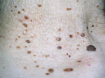
Seborrheic keratosis is an epidermal cutaneous neoplasm that carries no risk of malignant degeneration. It is, undoubtedly, one of the most common benign tumors of adults. However, although rare, it may also be seen in childhood. The number of lesions increases with age, and many elderly people have numerous tumors. Classic lesions of seborrheic keratosis (other than dermatosis papulosa nigra) are rare in dark-skinned individuals. The great majority of patients, including the ones with hundreds of lesions, are usually otherwise healthy. The trunk is the most common location ( see Fig. ) may sometimes be involved.
The clinical spectrum of seborrheic keratosis is wide. The lesions are observed in varying sizes, shapes, and colors. Although most lesions are 0.5 to 1.5 cm in diameter, tiny papules measuring 0.2 to 0.3 mm in diameter ( see Fig. ).
The lesions of seborrheic keratosis are typically well-circumscribed. Sometimes the border is notched ( see Fig. ).
A remarkably dark, flat, or thick seborrheic keratosis ( see Figs. ) may also be challenging in the differential diagnosis of various melanocytic tumors. This type of seborrheic keratosis has been called melanoacanthoma.
There are other clinical presentations of seborrheic keratosis, mainly related to its localization. Flat lesions may be encountered more commonly on the face ( see Fig. ).
Multiple seborrheic keratoses on the back may show a raindrop pattern, appearing as small, slightly elevated, linear lesions in a parallel distribution ( see Figs. ).
Although they usually cause no symptoms, some seborrheic keratoses may be pruritic. Inflammation caused by trauma of clothings or rupture of a horn cyst can cause erythema, erosion ( see Fig. ). Inflammation of irritated seborrheic keratoses regresses with the use of topical therapies in a few days; therefore there is no need for further treatment and prompt diagnostic intervention.
The Leser-Trlat sign is described as the sudden eruptive appearance and rapid increase in the size and number of seborrheic keratoses ( see Fig. ). It is considered a marker of internal malignancy, mainly adenocarcinoma of the gastrointestinal tract. However, the sudden development of numerous seborrheic keratoses only depends on the anamnesis, and most patients actually do not remember the exact time of onset of the lesions. A great majority of patients with numerous seborrheic keratoses are otherwise healthy. This is a very rare paraneoplastic sign. Therefore, a complete gastrointestinal evaluation of the patient, including endoscopic examination, is not commonly considered. It should be noted that pregnancy and erythroderma related with drug eruptions and inflammatory dermatoses like psoriasis and pityriasis rubra pilaris may also be associated with eruptive seborrheic keratoses.
Most seborrheic keratoses are diagnosed clinically but dermoscopy is also helpful. A punch biopsy may be indicated in lesions creating a diagnostic challenge. Histologically, seborrheic keratosis is mainly characterized by proliferation of small basaloid keratinocytes. The histopathologic appearance differs somewhat according to the type of the lesion. The most common hyperkeratotic type has a verruciform silhouette and a thickened keratin layer. In the acanthotic type, epidermal thickening, basaloid cell proliferation, and horn cysts (infoldings of surface keratin) are the main features. Irritated seborrheic keratosis is characterized by proliferation of larger keratinocytes resembling spinal layer cells, horn pearl formation, and an inflammatory infiltration. Differential diagnosis of this latter type from squamous cell carcinoma can sometimes be difficult. The exophytic structure and the presence of basaloid keratinocytes are helpful clues in favor of seborrheic keratosis.
Malignant tumors like basal cell carcinoma occuring within seborrheic keratosis is extremely rare and this seems to be an incidental phenomenon (see Fig. ).
Management . Seborrheic keratosis is a benign tumor. Unless a suspicion of Leser-Trlat sign is present, further examination is not necessary. Although the number of tumors may increase in time, individual lesions do not usually enlarge after they reach a certain size. Spontaneous regression is not expected but may occur in exceedingly rare cases. An accurate diagnosis is important to prevent unnecessary aggressive surgery that may result in unsightly scars. Many patients do not request therapy if they are fully informed about the benign nature of the tumor. However, therapy can be performed for cosmetic reasons or if lesions become troublesome. Cryotherapy, electrocautery, carbon dioxide laser, or Er: YAG laser ablation and simple curettage are therapeutic options that are usually cosmetically helpful. Shave excision may be preferred as a surgical method. However, all these procedures may result in postinflammatory hypo- or hyperpigmentation ( see Figs. ).
Although it is not seen very often, lesions may recur after therapy. Recurrent lesions occur overlying the intervention area or on the border as one or several sharply demarcated papules ( see Figs. ). Routine follow-up of seborrheic keratoses is not necessary, but in patients with multiple tumors, new occurrence of a different kind of pigmented or precancerous lesion may be confused with seborrheic keratosis, leading to a delay in consulting a dermatologist. Patients should be advised of the possibility of such a condition.