Nanette B. Silverberg Atlas of Pediatric Cutaneous Biodiversity 2012 Comparative Dermatologic Atlas of Pediatric Skin of All Colors 10.1007/978-1-4614-3564-8_1 Springer Science+Business Media, LLC 2012
1. The Appearance of Normal Skin
Nanette B. Silverberg 1 and Nanette B. Silverberg 2
(1)
Columbia University College of Physicians and Surgeons, 1090 Amsterdam Avenue, Suite 11D, New York, NY 10025, USA
(2)
Department of Dermatology, Pediatric and Adolescent Dermatology St. Lukes-Roosevelt Hospital and Beth Israel Medical Centers, 1090 Amsterdam Avenue, Suite 11D, New York, NY 10025, USA
Abstract
Table 1.1 reviews the standard Fitzpatrick skin types as they may apply to children by race and ethnicity. The table also includes potential skin issues that are commonly noted in my clinical practice, based on these skin tones, race, and ethnicity.
Fitzpatrick Phototyping Scale (Table )
Table reviews the standard Fitzpatrick skin types as they may apply to children by race and ethnicity. The table also includes potential skin issues that are commonly noted in my clinical practice, based on these skin tones, race, and ethnicity.
Table 1.1
Typical reaction patterns noted by race and ethnicity
Race/ethnicity | Fitzpatrick types | Skin reactions |
|---|
Asian/East Asian | IIIV | Risk of sun damage, dyspigmentation, facial sensitivity, and keloids |
Black/African American/Afro-Carribean | IVVI | Risk of dyspigmentation, especially Hyperpigmentation Obfuscation of diagnosis due to pigmentary alterations Xerosis Keloids Traction/styling hair damage |
Caucasian | I, II (rarely III) | Risk of phototoxic reactions, damage, and vascular reactivity in response to trauma and ultraviolet light exposure |
South east Asian/Indian | IIIV | Risk of pigmentary disturbance, especially hypopigmentation Xerosis |
Hispanic/Latino | IIV | Risk of dyspigmentation, both hyperpigmentation and hypopigmentation, obfuscation of diagnosis due to pigmentary alterations, xerosis, traction-related hair damage, keloids, and photodamage |
Native American | IIIV | Risk of dyspigmentation, both hyperpigmentation and hypopigmentation, xerosis, obfuscation of diagnosis due to pigmentary alterations and photodamage |
Middle Eastern | IIIVI | Risk of dyspigmentation, both hyperpigmentation and hypopigmentation, xerosis, obfuscation of diagnosis due to pigmentary alterations and photodamage |
Modified from Silverberg NB. Pediatric dermatology in children of color. Access Dermatol. 2010
Caucasian-Normal Skin Tone (Newborn) (Figs. 1.1 and 1.2)
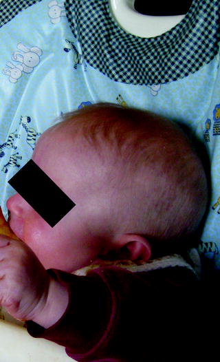
The normal skin of a newborn Caucasian child (Fig. 1.1) is lightly pigmented and can demonstrate light colored vellus hairs, prominent vasculature (Fig. 1.3), and easy formation of erythema with trauma. Furthermore, cutis marmorata can be prominent on the dependent portions of the body. Early changes that can be noted include milia, sebaceous hyperplasia, and infantile acne (). In early childhood, the skin loses much of the vascular reactivity patterns of infancy.
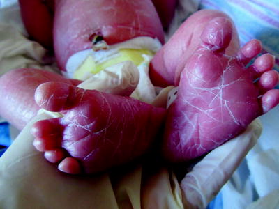
Caucasian-Postdates Peeling (Figs. 1.3 and 1.4)
Postdates peeling is common in children who are 40 or more weeks gestational age and can range from a branny to thick coating of shiny hyperkeratosis, resembling the thicker encasements of collodion membranes. Accompanying hyperlinearity of the palms and soles can be noted (Fig. 1.3).
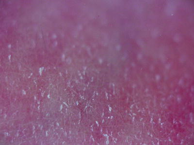
Dermoscopy demonstrates desquamation of the tiny stratum corneum keratinocytes around the edges (Fig. 1.4). Erythema noted in the background is a normal form of cutis marmorata of infancy, most obvious in Caucasian infants.
Hispanic/Latino-Normal Skin Tone (Newborn) (Figs. 1.5 and 1.6)
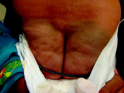
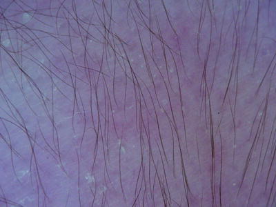
Fine lanugo and/or vellus hairs and mild desquamation of keratinocytes (Fig. 1.5), especially around follicular orifices, typifies newborn Hispanic/Latino skin. Lanugo hairs (Fig. 1.6) are noted in premature infants as they disappear towards the end of the third trimester, replaced by vellus hairs. Lanugo hairs and infantile vellus hairs are generally dark brown in this group of children as noted on the lower back in the Hispanic infant pictured in Figs. 1.5 and 1.6 (dermoscopic photo of the lower back seen in Fig. 1.5). Vellus hairs will usually disappear in the first year of life. However, some hirsute Hispanic/Latino children may have notable persistence of fine dark body hair over the trunk, back, and extremities through childhood. Congenital hairy pinna can be noted in infants of mothers with maternal gestational diabetes mellitus, but is also more common in individuals from the South Pacific, India, and Sri Lanka, as well as in some populations of Europe, Africa, and Australia [].
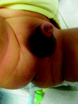
Black/African American Normal Skin Tone (Newborn) (Fig. 1.7)
In newborns of Black, African American or Afro-Caribbean descent hyperpigmentation can be noted over the ears, lips, fingertips, genitalia (Fig. 1.7) (especially in the midline), nipples, umbilicus, axillae, and anal orifice. Only axillary hyperpigmentation disappears by the end of the first year [).
Table 1.2
Ultrastructural differences in melanocyte distribution and melanosome packaging by race and ethnicity
Race | Pigmentary differences |
|---|
Black (in the United States: African American or Afro-Caribbean) | Large (stage IV), dispersed melanosomes |
Eumelanin |
Closely packed doublet or singlet melanosomes, rare aggregates []. Larger melanophages (May account in part for greater incidence of melasma and erythema Dyschromicum Perstans) |
UV filtration in the malpighian layer |
|

