Lidia Rudnicka , Malgorzata Olszewska and Adriana Rakowska (eds.) Atlas of Trichoscopy 2012 Dermoscopy in Hair and Scalp Disease 10.1007/978-1-4471-4486-1_1 Springer-Verlag London 2012
1. Introduction
Abstract
Trichoscopy may be performed with any dermoscope. This chapter focuses on the features of different types of dermoscopes, such as handheld dermoscopes, basic digital dermoscopes, dermoscopic accessories for the iPhone, and advanced digital dermoscopes (videodermoscopes). Basic information is provided on performing trichoscopy.
Any handheld dermoscope may be used to perform trichoscopy. Digital dermoscopes (videodermoscopes) also may be used. These have the advantages of easier photography and a higher magnification but are time consuming and expensive.
Among handheld dermoscopes, there are devices that require immersion fluid and those that use polarized light to cancel out reflections from the stratum corneum. Polarized light dermoscopes may have a contact or noncontact lens. Devices that combine contact and noncontact attributes (hybrid dermoscopes) also are available. The choice of a particular device is a matter of individual preference.
Results of studies addressing the differences among these three types of dermoscopes (nonpolarized light, polarized light, and noncontact polarized light) in skin cancer indicate that keratotic structures are visualized better in nonpolarized light, whereas blood vessels are seen best with noncontact polarized light dermoscopes []. Considering that most published trichoscopic images have been taken with nonpolarized light, this type of dermoscope may appear slightly more beneficial than polarized light dermoscopes for learning trichoscopy.
Most handheld dermoscopes allow one to observe the skin surface at a 10-fold magnification, whereas digital dermoscopes have working magnifications from 20- to 100-fold and higher. Lower magnifications have the benefit of providing a better overview of a large scalp area, whereas higher magnifications allow visualization of fine details. Most images in this atlas were taken at a 20- or 70-fold magnification (FotoFinder 2 dermoscope; FotoFinder Systems GmbH, Bad Birnbach, Germany).
Trichoscopy differs from skin cancer dermoscopy, among others, in the large area that needs to be examined. In diffuse hair loss, the frontal and occipital areas should be investigated. In cases of focal alopecia, the hairless plaque and hair-bearing margin should be examined. In some diseases, additional evaluation of the eyebrows may be informative.
When using a contact dermoscope, the roll-on technique of applying the lens to the scalp surface allows the best image analysis. In this technique, one edge of the lens is placed against the scalp surface at an approximately 45 angle, then the lens is rolled until the whole lens is in contact with the skin surface []. This technique allows detailed visualization of the structures when applying different amounts of pressure. When immersion fluid is used, this technique also allows one to squeeze out air inclusions (bubbles).
Nonpolarized light dermoscopes require immersion fluid (alcoholic, aqueous, or oily). Various types of immersion fluids exist: alcohol solutions, aqueous solutions, gels, and oils []. This method, called dry trichoscopy, allows better visualization of perifollicular scaling.
Hair washing generally does not influence trichoscopic results. Hair colorization also does not hinder trichoscopic analysis; it may even be beneficial for analyzing hair shaft structure and evaluating hair thickness in persons with light blond or gray hairshaft structure abnormality (pili torti).
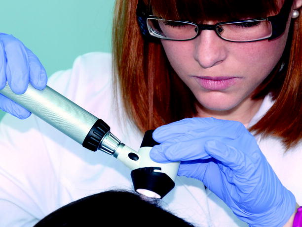
Fig. 1.1
Dr . Borkowska performing trichoscopy with a handheld dermoscope . Any handheld dermoscope may be used to perform trichoscopy. It does not require any additional lenses or parts. You may use your dermoscope just as you would for diagnosing melanoma
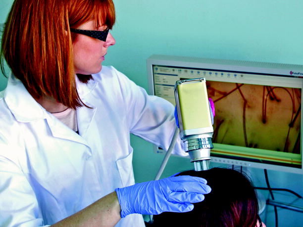
Fig. 1.2
Dr . Borkowska performing trichoscopy with a digital dermoscope ( videodermoscope ). A digital dermoscope (videodermoscope) has the benefit of better working comfort, higher magnification, and easy photography. It is, however, more time consuming and expensive. Thus, it rarely is used in clinical practice. The old term videodermoscopy refers to dermoscopy performed with a digital dermoscope
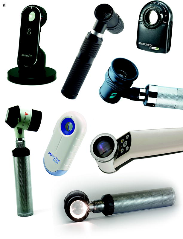
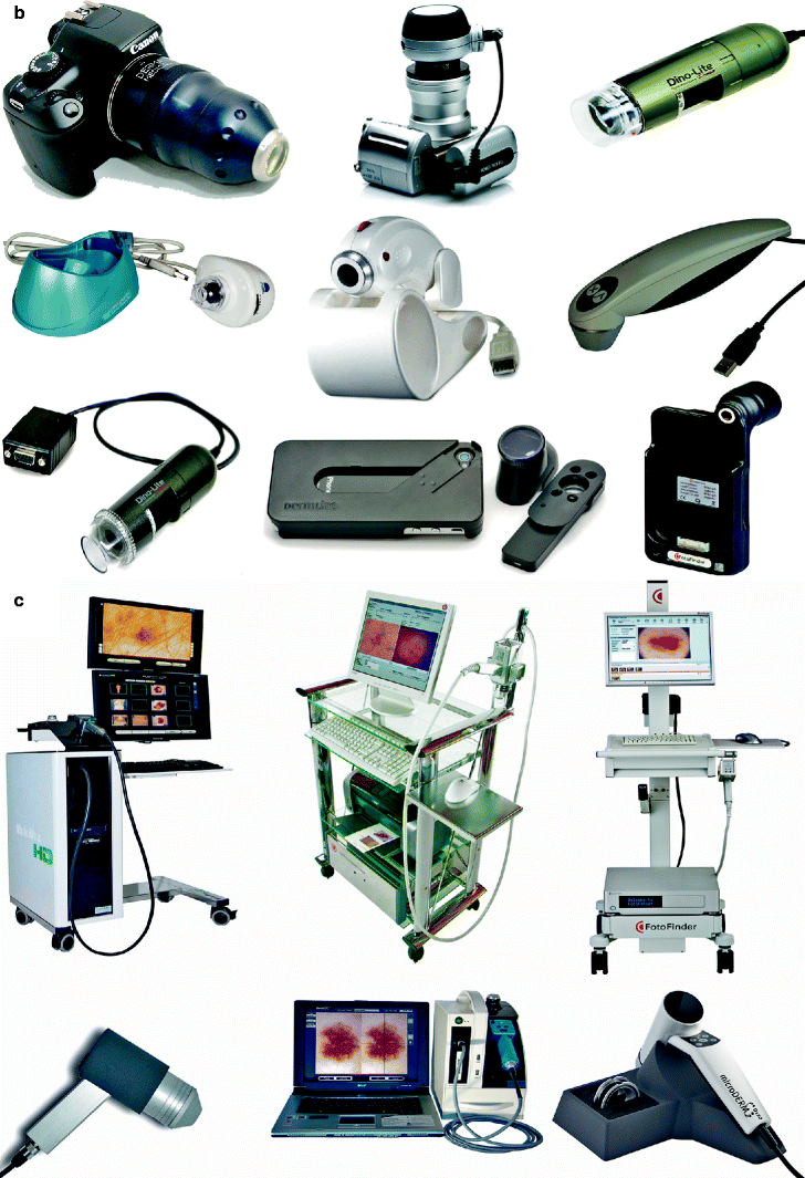
Fig. 1.3
Examples of equipment that may be used to perform trichoscopy . Trichoscopy may be performed with ( a ) handheld dermoscopes, ( b ) basic digital dermoscopes and photographic equipment, or ( c ) advanced digital dermoscopes. Handheld dermoscopes may be divided into three groups: contact dermoscopes, polarized light contact dermoscopes, and polarized light noncontact dermoscopes. Also available are handheld dermoscopes that work in either the contact or noncontact mode; these are known as hybrid dermoscopes. Which device to choose is a matter of individual preference; there is no preferred type of dermoscope for performing hair and scalp examinations. The standard magnification of handheld dermoscopes is 10; the cost varies between about US $700 and US $1,800 ( a ). New devices on the market include simplified digital dermoscopes that may be connected to a computer (e.g., via USB) and kits allowing one to connect selected handheld dermoscopes to a regular photo camera or to an iPhone 4/4S. They are a good solution for dermatologists who like to take trichoscopic photographs in their daily practice. The usual magnification is 10 to 80, depending on the device. Although higher magnifications may be possible, they usually are not sufficiently supported by the light source of the equipment currently available. The price of these devices varies between US $400 and US $2,000 (not including the computer, camera, or iPhone) ( b ). The large, expensive digital dermoscopes (videodermoscopes) allow one to take high-magnification, high-quality photographs. The price of these devices varies significantly, depending on the presence or absence of software, which is not necessary for trichoscopy. Most companies now offer these devices with laptops instead of full-size computers. This type of digital dermoscope offers multiple magnifications in the range of 20 to 70 (or 100) and higher. The price varies from about US $10,000 to about US $20,000 ( c ). These images show examples of dermoscopes available on the market when we were writing this chapter ( Courtesy of 3Gen LLC, San Juan Capistrano, CA; AnMo Electronics Corp., New Taipei City, Taiwan; Bechtold & Co, Lodz, Poland; Consultronix S.A., Krakow, Poland; Delasco Dermatologic Lab & Supply, Inc, Council Bluffs, IA; Derma Medical Systems GmbH, Vienna, Austria; DermoScan GmbH, Regensburg, Germany; FotoFinder Systems GmbH; Medit Inc., Winnipeg, MB, Canada; IDCP B.V., Naarden, The Netherlands; Schuco International Ltd., London, UK.)

