Part 1
Dysphagia
George Y. Wu , Nathan Selsky and Jane M. Grant-Kels (eds.) Atlas of Dermatological Manifestations of Gastrointestinal Disease 2013 10.1007/978-1-4614-6191-3_1 Springer Science+Business Media New York 2013
1. Oropharyngeal Cancer: Gastrointestinal Features
Laura Nieves 1
(1)
Department of Internal Medicine, University of Connecticut Health Center, Farmington, CT, USA
Abstract
Oropharyngeal cancer comprises the malignant pathologies involving the tissues of the oropharynx. This includes the tongue, tonsils, soft palate, and walls of the pharynx. These cancers are generally divided into those that are human papillomavirus (HPV)-positive and those that HPV-negative (generally related to smoking or alcohol).
Oropharyngeal cancer comprises the malignant pathologies involving the tissues of the oropharynx. This includes the tongue, tonsils, soft palate, and walls of the pharynx. These cancers are generally divided into those that are human papillomavirus (HPV)-positive and those HPV-negative (generally related to smoking or alcohol). The gastrointestinal (GI) symptoms include []:
The most frequent clinical signs and findings are []:
Nonhealing sores or ulcers in oral cavity (pain or painless)
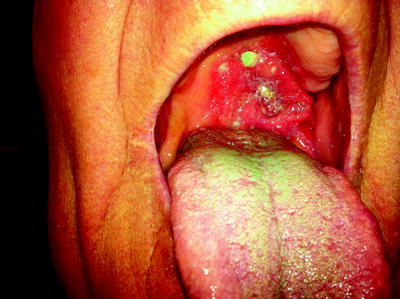
Red or white lesion with an irregular, fungating growth; surface ulcerations; easy bleeding ( see Fig. )
Cervical lymphadenopathy
Associated conditions:
Bazexs syndrome (acrokeratosis paraneoplastica), rare, erythematous to violaceous psoriasiform plaques in acral areas. Palmoplantar keratoderma, alopecia, and nail dystrophy are common. Has also been associated with other malignancies.
Fig. 1.1
Extensive oropharyngeal carcinoma associated with Bazexs syndrome
The pathogenesis involves the usual risk factors for upper GI malignancy []:
Chronic inflammation (thought to be precipitating factor)
Series of somatic or epigenetic changes allow resistance to growth-inhibitory signals, autonomous proliferation, avoidance of apoptosis, unlimited replication, angiogenesis, invasion and metastasis.
The pathology shows typical features of mucosal malignancy []:
The diagnosis is made by considering the following []:
Initial assessment by careful examination of oral cavity
Confirmed by biopsy
Panendoscopy (including laryngoscopy, esophagoscopy, and possible bronchoscopy) looking for other head and neck tumors
Magnetic resonance imaging (MRI), computed tomography (CT) scan, and positron emission tomography (PET) scan for staging
The differential diagnosis of oropharyngeal cancer should include []:
The treatment involves usual modalities for the treatment of malignancy [6]:
Multimodaldepends on staging and location
Principal treatment modalities are surgery and radiation therapy (RT)
Post-surgical chemoradiation (platinum-based) for advanced disease
Radiotherapy plus cetuximab has survival benefit over RT alone
Cessation of risk factors (alcohol and tobacco)
References
Moore RL, Devere TS. Epidermal manifestations of internal malignancy. Dermatol Clin. 2008;26:1729. PubMed CrossRef
European Society for Medical Oncology (ESMO). Squamous cell carcinoma of the head and neck: ESMO Clinical Recommendations for diagnosis, treatment and follow-up. Ann Oncol. 2007;18:ii656. CrossRef
Kupferman ME, Myers JN. Molecular biology of oral cavity squamous cell carcinoma. Otolaryngol Clin N Am. 2006;39:22934. CrossRef
Greer R. Pathology of malignant and premalignant oral epithelial lesions. Otolaryngol Clin N Am. 2006;39:24975. CrossRef
Chen AY. Cancer of the oral cavity. Dis Mon. 2001;47:275361. PubMed CrossRef
Scottish Intercollegiate Guidelines Network (SIGN). Diagnosis and management of head and neck cancer. A national clinical guideline. Edinburgh, Scotland): Scottish Intercollegiate Guidelines Network (SIGN); 2006
George Y. Wu , Nathan Selsky and Jane M. Grant-Kels (eds.) Atlas of Dermatological Manifestations of Gastrointestinal Disease 2013 10.1007/978-1-4614-6191-3_2 Springer Science+Business Media New York 2013
2. Bazexs Syndrome: Dermatological Features (Acrokeratosis Paraneoplastica)
Liam Zakko 1
(1)
Yale Department of Internal Medicine, Yale New Haven Hospital, 20 York Street, New Haven, CT 06510, USA
(2)
Department of Dermatology, University of Connecticut Health Center, 21 South Road, Farmington, CT 06030, USA
Liam Zakko (Corresponding author)
Email:
Abstract
Clinical signs and features include:
Paraneoplastic syndrome characterized by acral psoriasiform lesions: red violaceous hyperkeratotic plaques with ill-defined borders; nail changes including subungual hyperkeratosis, onycholysis, longitudinal streaks, yellow pigmentation; hyperkeratotic plaques at pressure points (knees, elbows); and hyperkeratotic plaques on the ears, nose, and cheeks
Particular findings include violet discoloration and bulbous enlargement of the distal phalanges; lesions only on the helix of the ear (not entire ear like psoriasis)
Three clinical stages: (1) poorly circumscribed skin lesions on the helices of the ears, nose, cheeks, fingers, toes, and nails associated with asymptomatic carcinomas most commonly of the upper aerodigestive tract ( see Fig. 2.1b); (2) lesions which spread to the palms and soles with a locally symptomatic malignancy ( see Fig. 2.1a); (3) With an untreated malignancy, erythema and scale spread to the knees, elbows, and trunk
Clinical signs and features include:
Paraneoplastic syndrome characterized by acral psoriasiform lesions: red violaceous hyperkeratotic plaques with ill-defined borders; nail changes including subungual hyperkeratosis, onycholysis, longitudinal streaks, yellow pigmentation; hyperkeratotic plaques at pressure points (knees, elbows); and hyperkeratotic plaques on the ears, nose, and cheeks []

