Part 1
Fundamentals
Alberto Rosenblatt , Homero Gustavo de Campos Guidi and Walter Belda Jr. Male Genital Lesions 2012 The Urological Perspective 10.1007/978-3-642-29017-6_1 Springer-Verlag Berlin Heidelberg 2013
1. Embryology and Functional Anatomy of the Male External Genitalia
Alberto Rosenblatt 1
(1)
Albert Einstein Jewish Hospital (HIAE), Av. Albert Einstein 627/701, 05651-901 So Paulo, SP, Brazil
(2)
University of So Paulo Medical School, Av. Eneas Carvalho de Aguiar, 255 - sl. 10.166, So Paulo, SP, Brazil
(3)
Department of Dermatology, University of So Paulo Medical School, Avenida Aoc 162, 04075020 So Paulo, SP, Brazil
Alberto Rosenblatt (Corresponding author)
Email:
Homero Gustavo de Campos Guidi
Email:
Abstract
Dermatoses of the male external genitalia may comprise either specific disorders of the genital region or local manifestations of systemic diseases. In addition, normal anatomical variations of the penis and scrotum are commonly observed, which may further confound the correct diagnosis of the innumerous conditions affecting the region. Therefore, understanding the basic notions of the male genital embryology and functional anatomy, as well as the correct sequence of history taking and physical examination are of utmost importance in order to better characterize the conditions involving this particular site. The embryology of the male external genitalia and its functional anatomy are discussed in this Chapter.
1.1 Introduction
Dermatoses of the male external genitalia may comprise either specific disorders of the genital region or local manifestations of systemic diseases. In addition, normal anatomical variations of the penis and scrotum are commonly observed, which may further confound the correct diagnosis of the innumerous conditions affecting the region. Therefore, understanding the basic notions of the male genital embryology and functional anatomy, as well as the correct sequence of history taking and physical examination are of utmost importance in order to better characterize the conditions involving this particular site. The embryology of the male external genitalia and its functional anatomy are discussed in this Chapter.
1.2 Embryology of the Male External Genitalia
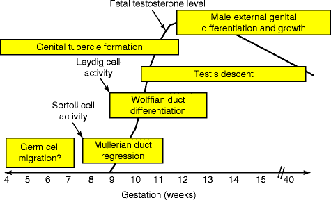
Fetal sex development involves a series of sequential stages (Fig. ).
Fig. 1.1
External genitalia differentiation/formation is a late developmental step during human ontogeny ( Source : Klonisch et al. (). Reproduced with permission from Elsevier)
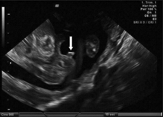
Fig. 1.2
Male genitalia at 15 weeks: the genital tubercle is pointing cranially ( arrow ) ( Source : Odeh et al. (). Reproduced with permission from Lippincott Williams & Wilkins)
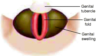
Fig. 1.3
Fetal sex development undifferentiated structures at 7 weeks of gestation
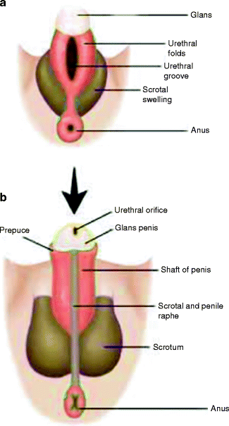
Fig. 1.4
Fetal sex development male external genital structures at 78 weeks ( a ) and 12 weeks of gestation ( b )
Fig. 1.5
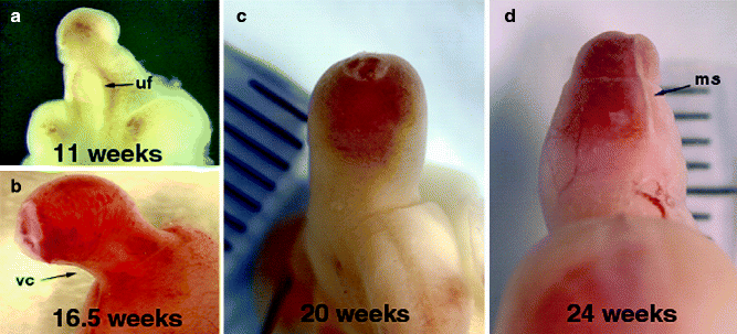
Male human fetal external genitalia during gestation. ( a ) 11 weeks, note the urethra is open and urethral fold ( uf ) and groove are prominent in the transillumination view of the phallus. ( b ) At 16.5 weeks, note the normal ventral curvature ( vc ) is shown and the foreskin which is almost completely formed. ( c ) At 20 weeks gestation penile and urethral development are complete with the prepuce covering the glans and the penile curvature resolved. ( d ) At 24 weeks the prepuce covers the whole glans, note the midline seam ( ms ). Note the progression of natural curvature to a straight phallus during development ( Source : Baskin (). Reproduced with permission from Elsevier)
Fig. 1.6
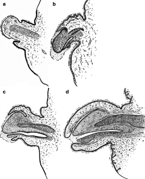
Longitudinal section (LS) of the penis of a human fetus: ( a ) with 40 mm Crown-Rump (C.R.) length. Note the ridge of ectoderm behind the corona and the glans, and the thin layer of desquamating cells on the surface. ( b ) With 70 mm. C.R. length. Note the dorsal ectodermal tissues thrown into loose folds, and the distribution of the desquamating cells. ( c ) With 100 mm. C.R. length. The prepuce is here seen overflowing the glans and enclosing a layer of desquamating cells between it and the glans. ( d ) With 170 mm. C.R. length. This section shows the prepuce covering the whole of the glans, with the layer of desquamating cells connecting the two. Note the plug of desquamating cells closing the opening of the urethra ( Source : Hunter ())
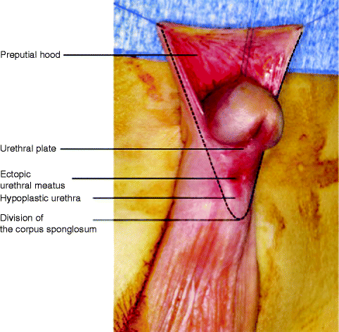
Cryptorchidism (the failure of the testes to descent into the scrotal sac) and hypospadias (where a cleft on the ventral aspect of the penis develops due to urethral tube dysmorphogenesis) (Figs. ).
Fig. 1.7
Genital abnormalities hypospadias with a midshaft division of the corpus spongiosum ( Source : Vidal et al. (). Reproduced with permission from Elsevier)
Fig. 1.8
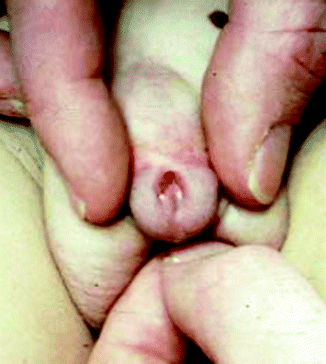
Genital abnormalities disorder of sex development (46,XY) showing severe hypospadias in partial androgen insensitivity ( Source : Woodward and Patwardhan (). Reproduced with permission from Elsevier)
Fig. 1.9
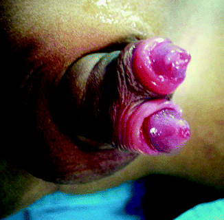
An epispadiac urethra in an otherwise normal penis ( Source : Woodhouse (). Reproduced with permission from Springer)
Fig. 1.10
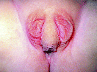
Genital abnormalities virilized external genitalia in a congenital adrenal hyperplasia patient with a large genital tubercle and two scrotalized genital folds ( Source : Vidal et al. (). Reproduced with permission from Elsevier)

