• Complain

Néstor P. Sánchez (editor) - Atlas of Dermatology in Internal Medicine

Here you can read online Néstor P. Sánchez (editor) - Atlas of Dermatology in Internal Medicine full text of the book (entire story) in english for free. Download pdf and epub, get meaning, cover and reviews about this ebook. year: 2011, publisher: Springer, genre: Home and family. Description of the work, (preface) as well as reviews are available. Best literature library LitArk.com created for fans of good reading and offers a wide selection of genres:

Romance novel Science fiction Adventure Detective Science History Home and family Prose Art Politics Computer Non-fiction Religion Business Children Humor

Choose a favorite category and find really read worthwhile books. Enjoy immersion in the world of imagination, feel the emotions of the characters or learn something new for yourself, make an fascinating discovery.

Néstor P. Sánchez (editor) Atlas of Dermatology in Internal Medicine

Atlas of Dermatology in Internal Medicine: summary, description and annotation

We offer to read an annotation, description, summary or preface (depends on what the author of the book "Atlas of Dermatology in Internal Medicine" wrote himself). If you haven't found the necessary information about the book — write in the comments, we will try to find it.

Featuring more than 150 clinical photographs, this is the only concise text-atlas on the most common and important cutaneous manifestations of systemic disease in both children and adults. It also includes a dedicated chapter on skin diseases seen in the ICU.

Néstor P. Sánchez (editor): author's other books


Who wrote Atlas of Dermatology in Internal Medicine? Find out the surname, the name of the author of the book and a list of all author's works by series.

Atlas of Dermatology in Internal Medicine — read online for free the complete book (whole text) full work

Below is the text of the book, divided by pages. System saving the place of the last page read, allows you to conveniently read the book "Atlas of Dermatology in Internal Medicine" online for free, without having to search again every time where you left off. Put a bookmark, and you can go to the page where you finished reading at any time.

Light

Font size:

Reset

Interval:

Bookmark:

Make
Nstor P. Snchez (ed.) Atlas of Dermatology in Internal Medicine 10.1007/978-1-4614-0688-4_1 Springer Science+Business Media, LLC 2012
Cutaneous Manifestations of Connective Tissue Diseases
Lesliane E. Castro-Santana 1 , Jos Gonzlez Chvez 2, Jennifer Rulln 2 and Rachelle E. Seijo-Montes 2
(1)
Department of Rheumatology, University of Puerto Rico School of Medicine, Medical Sciences Campus, San Juan, Puerto Rico
(2)
Department of Dermatology, University of Puerto Rico School of Medicine, Medical Sciences Campus, San Juan, Puerto Rico
Lesliane E. Castro-Santana
Email:
Abstract
Most connective tissue diseases are characterized by a diversity of systemic and cutaneous manifestations. In this chapter, we focus on the unique cutaneous manifestations that characterize systemic lupus erythematosus, dermatomyositis, and scleroderma. Observation and histopathologic evaluation of the skin can assist the clinician in initiating therapy in a timely manner, even before the onset of systemic disease. A detailed discussion of these cutaneous and histopathological manifestations is presented.
Introduction
Lupus erythematosus, dermatomyositis, scleroderma, and rheumatoid arthritis are connective tissue diseases characterized by a diversity of systemic and cutaneous manifestations. In some cases, the skin changes may precede the development of overt disease and may, in fact, be the only indicator of the patients disease []. It is important to recognize these signs early to institute adequate and timely therapy.
Systemic Lupus Erythematosus
Systemic lupus erythematosus (SLE) is an autoimmune inflammatory disease, which can affect multiple organ systems, including the skin []. These will each be discussed in detail in the remainder of the chapter.
Acute Cutaneous Lupus Erythematosus
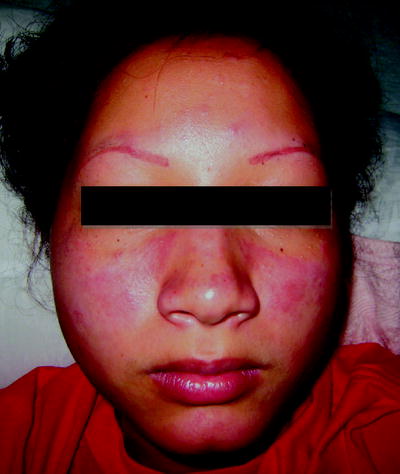
Malar erythema is commonly the first manifestation of SLE, and the most common manifestation of acute cutaneous lupus erythematosus (ACLE). It can precede other systemic manifestations of SLE by weeks or months, and frequently coincides with disease exacerbations. The rash is characterized by erythema, edema, and occasionally fine scales in a malar distribution, along the bridge of the nose, with characteristic sparing of the nasolabial folds (Fig. ].
Fig 1 Acute cutaneous lupus erythematosus associated with systemic lupus - photo 1
Fig. 1
Acute cutaneous lupus erythematosus associated with systemic lupus erythematosus
Subacute Cutaneous Lupus Erythematosus
Subacute cutaneous lupus erythematosus (SCLE) is a distinctive subset of lupus erythematosus. Patients can develop cutaneous lesions with two different morphologies: papulosquamous and annular, polycyclic lesions. There is no induration or telangiectasias. The distribution of the lesions is generally on sun-exposed areas, particularly the dorsal surface of upper extremities, upper back, and chest [].
Histopathologically, the lesions are very similar to those of discoid lupus erythematosus (DLE); however, SCLE lesions have minimal hyperkeratosis and follicular plugging. Instead, they are characterized by more prominent epidermal atrophy and liquefaction degeneration of the basal cell layer. The inflammatory infiltrate in contradistinction to DLE tends to be scattered throughout the reticular dermis, but similar to DLE, is composed of activated T-cells [].
Chronic Cutaneous Lupus Erythematosus
The wide spectrum of CCLE includes the classical discoid lesions that can be localized or generalized, chilblain, hypertrophic, verrucous, lichenoid, lupus panniculitis (profundus), mucosal discoid, palmoplantar erosive, or lupus erythematosus tumidus lesions []. We will discuss the most commonly seen.
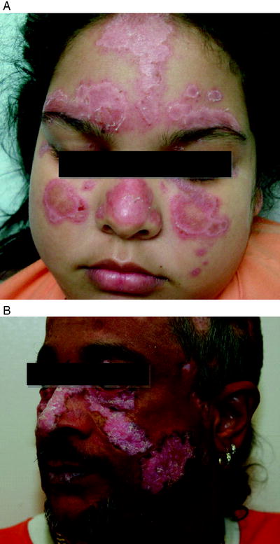
The lesions of DLE are characterized by well-defined, disk-shaped, erythematous plaques with epidermal atrophy, telangiectasias, and scaling (Fig. ].
Fig 2 a b Discoid lesions of lupus erythematosus chronic cutaneous - photo 2
Fig. 2
( a , b ) Discoid lesions of lupus erythematosus (chronic cutaneous lupus erythematosus)
Hypertrophic DLE is a distinct form of DLE, which is characterized by hyperkeratotic plaques that typically are observed over the face, extensor surfaces of the arms, legs, and upper trunk [].
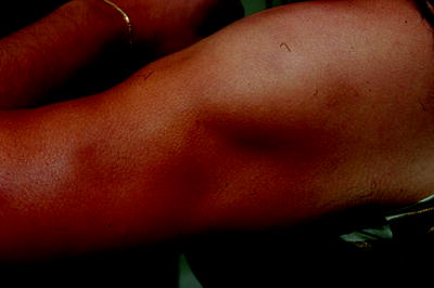
Lupus panniculitis (also known as lupus profundus) is a condition characterized by multiple firm, well-demarcated subcutaneous nodules or plaques that are persistent and painless (Fig. ].
Fig 3 Lupus panniculitis lupus profundus Well-demarcated depressed plaques - photo 3
Fig. 3
Lupus panniculitis (lupus profundus): Well-demarcated depressed plaques with atrophy located on the upper extremity of a patient with lupus panniculitis
Chilblain lupus is a rare manifestation of CCLE consisting of lesions induced by cold in acral areas. They are pruritic or painful and can have erythematous papules that ulcerate or become hyperkeratotic [].
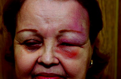
Lupus erythematosus tumidus is characterized by marked photosensitivity and a tendency to occur in the male sex (Fig. ].
Fig 4 Lupus erythematosus tumidus Succulent erythematous plaque involving - photo 4
Fig. 4
Lupus erythematosus tumidus. Succulent, erythematous plaque involving left upper forehead
The differential diagnosis of cutaneous lupus erythematosus includes sunburn, rosacea, seborrheic dermatitis, allergic contact dermatitis, phototoxic drug eruption, polymorphous light eruption, tinea faciei, granuloma faciale, sarcoidosis, alopecia mucinosa, and benign lymphocytic infiltrate of Jessner [].
Bullous Systemic Lupus Erythematosus
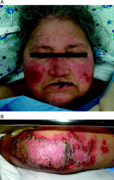
The bullous lesions of lupus erythematosus are predominantly located on the face, neck, and upper trunk. They tend to be tense and may rupture leaving erosions, crusts, and changes in pigmentation (Fig. ].
Fig 5 a b Exacerbation of systemic lupus erythematosus presenting - photo 5
Fig. 5
( a , b ) Exacerbation of systemic lupus erythematosus presenting clinically with bullaes and erosions in skin and mucous membranes. Blistering usually indicate a more severe systemic disease
Classification and Treatment
The American College of Rheumatology has developed criteria for the classification of SLE (Table ].
Table 1
ACR criteria for the classification of systemic lupus erythematosus
1.Malar rash
2.Discoid rash
3.Photosensitivity (by patient history or physician observation)
Next page
Light

Font size:

Reset

Interval:

Bookmark:

Make

Similar books «Atlas of Dermatology in Internal Medicine»

Look at similar books to Atlas of Dermatology in Internal Medicine. We have selected literature similar in name and meaning in the hope of providing readers with more options to find new, interesting, not yet read works.


Reviews about «Atlas of Dermatology in Internal Medicine»

Discussion, reviews of the book Atlas of Dermatology in Internal Medicine and just readers' own opinions. Leave your comments, write what you think about the work, its meaning or the main characters. Specify what exactly you liked and what you didn't like, and why you think so.