Ali Hendi and Juan Carlos Martinez Atlas of Skin Cancers Practical Guide to Diagnosis and Treatment 10.1007/978-3-642-13399-2_1 Mayo Foundation for Medical Education and Research 2011
1. Introduction
Ali Hendi 1, 2, 3
(1)
Department of Dermatology, Mayo Clinic, Jacksonville, FL, USA
(2)
College of Medicine, Mayo Clinic, Rochester, MN, USA
(3)
Private Practice, Chevy Chase, MD, USA
Ali Hendi Consultant, Assistant Professor of Dermatology (Corresponding author)
Email:
Juan-Carlos Martinez Senior Associate Consultant, Assistant Professor of Dermatology
Email:
Abstract
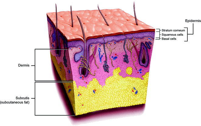
The skin is the largest organ of the body. A basic understanding of the anatomy of the skin is needed for the proper diagnosis and management of skin cancers. The basic subunits of the skin include the epidermis, dermis, and subcutis (Fig. 1.1). The epidermis is the outermost layer of the skin. The innermost layer of the epidermis is the basal layer. The basal layer cells multiply to form the squamous layer of the epidermis. The outer layer of epidermis is the stratum corneum, which is composed of keratin. Melanocytes (pigment-producing cells) lie within the basal layer of the epidermis. The dermis incorporates the adnexal structures (hair follicles, sebaceous glands, and sweat glands) and is made up of collagen, elastic tissue, and reticular fibers. The subcutis is made up of adipose tissue.
The skin is the largest organ of the body. A basic understanding of the anatomy of the skin is needed for the proper diagnosis and management of skin cancers. The basic subunits of the skin include the epidermis, dermis, and subcutis (Fig. ). The epidermis is the outermost layer of the skin. The innermost layer of the epidermis is the basal layer. The basal layer cells multiply to form the squamous layer of the epidermis. The outer layer of epidermis is the stratum corneum, which is composed of keratin. Melanocytes (pigment-producing cells) lie within the basal layer of the epidermis. The dermis incorporates the adnexal structures (hair follicles, sebaceous glands, and sweat glands) and is made up of collagen, elastic tissue, and reticular fibers. The subcutis is made up of adipose tissue.
Fig. 1.1
Schematic detailing the multiple layers of the skin
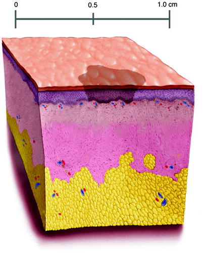
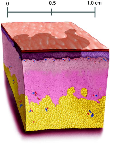
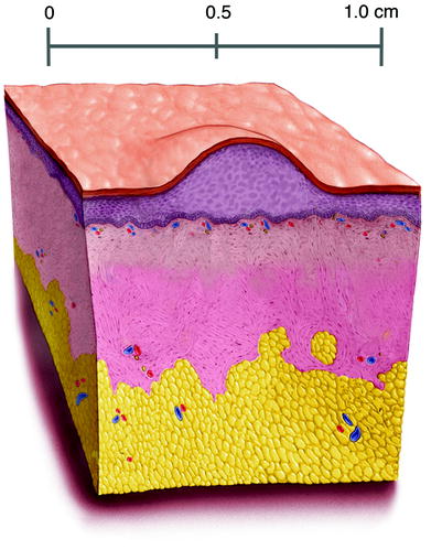
There are terms used in describing skin lesions that are unique to dermatology. This terminology is helpful in documenting lesions and in communicating with colleagues. The basic terminology and associated illustrations are explained in Figs..
Fig. 1.2
Macule-Flat, <5 mm, circumscribed skin discoloration that lacks surface elevation or depression
Fig. 1.3
Patch-Flat, >5 mm, circumscribed skin discoloration; a large macule
Fig. 1.4
Papule-Elevated, solid lesion, <0.5 cm in diameter
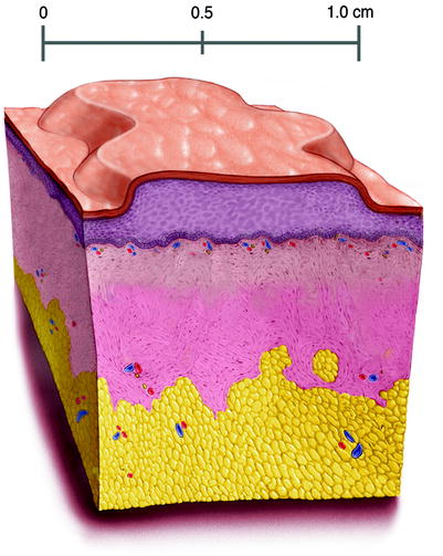
Fig. 1.5
Plaque-Elevated, flat-topped, solid lesion, >0.5 cm in diameter
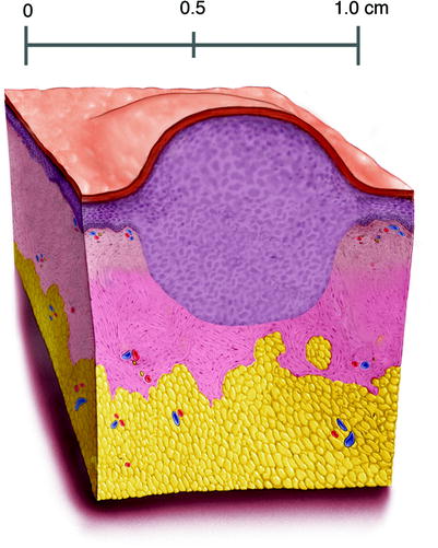
Fig. 1.6
Nodule-Elevated, circular or domed, solid lesion; >0.5 cm in diameter; a large papule
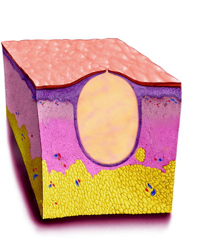
Fig. 1.7
Cyst, a well-circumscribed nodule that has an epithelial lining; generally has a punctum or connection to the surface
Ali Hendi and Juan Carlos Martinez Atlas of Skin Cancers Practical Guide to Diagnosis and Treatment 10.1007/978-3-642-13399-2_2 Mayo Foundation for Medical Education and Research 2011
2. Actinic Keratosis
Ali Hendi 1, 2, 3
(1)
Department of Dermatology, Mayo Clinic, Jacksonville, FL, USA
(2)
College of Medicine, Mayo Clinic, Rochester, MN, USA
(3)
Private Practice, Chevy Chase, MD, USA
Ali Hendi Consultant, Assistant Professor of Dermatology (Corresponding author)
Email:
Juan-Carlos Martinez Senior Associate Consultant, Assistant Professor of Dermatology
Email:
Abstract
While actinic keratoses (AKs) are commonly considered to represent a premalignant condition, some authors have asserted that these, in fact, represent the earliest stage of squamous cell carcinoma (SCC) (Ackerman and Mones 2006). Nevertheless, the majority of clinicians do not treat these as malignancies but as precursor lesions with malignant potential. Very common in older, photodamaged patients, they appear as gritty, scaly, pink plaques that are often easier to feel than see. For this reason, palpation of the skin is important in their detection.
2.1 Introduction
While actinic keratoses (AKs) are commonly considered to represent a premalignant condition, some authors have asserted that these, in fact, represent the earliest stage of squamous cell carcinoma (SCC) (Ackerman and Mones ). Nevertheless, the majority of clinicians do not treat these as malignancies but as precursor lesions with malignant potential. Very common in older, photodamaged patients, they appear as gritty, scaly, pink plaques that are often easier to feel than see. For this reason, palpation of the skin is important in their detection.
Actinic keratoses that become persistent or hypertrophic may evolve into squamous cell carcinoma, and, therefore, they are usually treated. Diagnosis is most commonly made based on clinical appearance. Questionable lesions, or those that fail to respond to initial treatments, should be biopsied to rule out squamous cell carcinoma.
2.2 Treatment of Actinic Keratoses
There are a number of methods available to satisfactorily treat actinic keratoses. Many actinic keratoses will spontaneously clear, and it is not unreasonable to manage conservatively with observation. This modality is best suited for reliable patients that are seen on a routine basis. As the progression of AK to SCC tends to occur rather slowly, observation may be well suited for patients with a decreased life expectancy. While the focus of this atlas is on skin cancer, for the sake of completeness, some of the most commonly used treatment methods for actinic keratoses are covered below.
2.2.1 Cryotherapy
Cryotherapy is most commonly used for the treatment of actinic keratoses. This technique usually involves the use of a specialized device that can very precisely control the flow of liquid nitrogen. It is important to recognize that while this technique can be very effective in the destruction of epidermal lesions, it is commonly accompanied by blistering, erosion, and can frequently result in a hypopigmented or depressed scar. Therefore, great care must be taken in limiting the amount of damage to the surrounding tissue, especially in more cosmetically sensitive areas such as the face.

