Armand B. Cognetta Jr. and William M. Mendenhall (eds.) Radiation Therapy for Skin Cancer 2013 10.1007/978-1-4614-6986-5_1 Springer Science+Business Media New York 2013
Abstract
Shortly after the discovery of X-rays in 1895, skin tumors and diseases were treated with the newly discovered form of ionizing radiation. As the X-ray tubes became more reliable, tissue response and fractionation became better understood and more effective regimens with fewer side effects were demonstrated. Superficial radiotherapy waned in popularity among dermatologists with the eventual arrival of topical steroids and Mohs surgery. In the last decade, however, there have been signs of resurgence among dermatologists who appreciate its utility in successfully treating skin cancers in an increasingly frail and elderly population in a nonsurgical manner with satisfactory cosmetic results.
On November 8, 1895, a German physicist and professor named Wilhelm Roentgen conducted some experiments with a cathode ray tube that led him to discover eine neue Art von Strahlena new kind of rays []. He chose the term X- strahlen X-rays as the type of rays because the frequency and characteristics of these rays were unknown. He had made the discovery when observing that the invisible cathode rays caused a fluorescent effect on a small cardboard screen painted with barium platinocyanide. The intensity of the fluorescence was diminished proportionally by distances and by certain filter materials of various thicknesses. Roentgen was later awarded the first Nobel Prize for physics in 1901 for his efforts and a bustling new era of applied physical science was spawned from the discovery and development of the X-rays.
The discovery of X-rays also generated interest in natural sources of radiation such as the study of visibly fluorescent compounds. In 1896 Becquerel discovered that radiation was naturally occurring in all uranium compounds. After an initially unsuccessful attempt to induce fluorescence, Becquerel placed the uranium salts and the photographic plates used in his experiments in a drawer with plans to recommence the experiment at a later date. Months later when he developed the photographic plate, he discovered a darkened area which could only be explained by something intrinsic to the uranium salts. Maria Curie furthered this research by testing various materials and discovered that compounds containing thorium also exhibited radioactive properties. Pierre Curie and his brother Paul-Jacques Curie subsequently created a device known as the piezoelectrometer which allowed study of the intensity of radioactive emissions. While studying the substance, pitchblende, Marie and her husband Pierre Curie discovered emissions that were too intense to be explained by the amount of known uranium in the substance. Through careful experimentation, they isolated a new element polonium (after Marias home country) and soon after came the discovery of radium. The Nobel Prize for physics was later awarded to Becquerel, Maria, and Pierre [].
The similarity of radiums effect on the skin to that of the X-ray was noted in 1901 by Pierre Curie and Becquerel following the work of German scientists Giesel and Walkoff which ultimately gave rise to Brachytherapy (from the Greek word brachys, meaning short). Brachytherapy involves the placement of the radiation source inside or in short proximity to the lesion or skin condition to be treated. A more detailed history of Brachytherapy can be found at the beginning of .
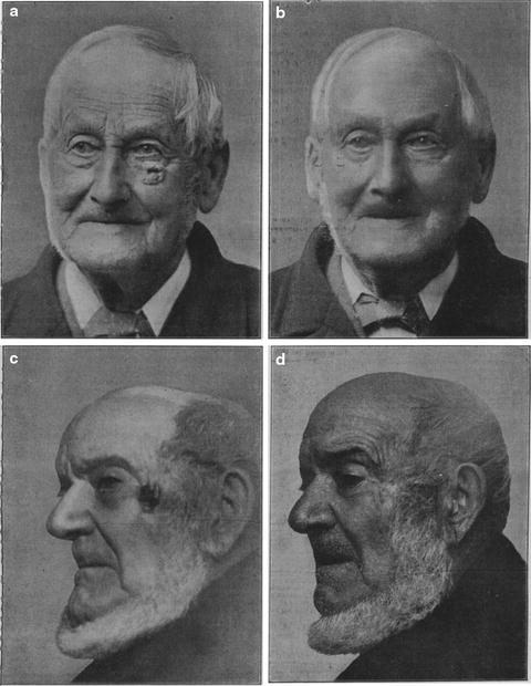
Less than a year after the discovery, X-rays began to be used in the treatment of skin disease. The first reported use of X-rays for the treatment of a disease of the skin was done by Leopold Fruend of Vienna in 1896 on a nevus pigmentosus piliferus located on the back of a 5-year-old girl. Multiple reports surfaced describing the efficacy of X-rays in the treatment of skin cancers, including J.W. Pughs article in 1902 entitled, Four Cases of Rodent Ulcer Treated by X Rays, in which before- and after-photographs were displayed [). With multiple early reports of success treating skin cancers with X-rays and a tremendous enthusiasm for its potential, Pusey, an American dermatologist attempted to formulate an appropriate therapeutic window for this new, powerful, and potentially dangerous modality in his lecture entitled Rationale of and the Indications for Therapeutic Use of Rontgen Rays, given at the 27th Annual Meeting of the American Dermatology Association in Washington on May 13th and 14th, 1903. This new treatment modality proved to be a tremendous dermatologic breakthrough affording success in treating numerous previously recalcitrant skin cancers and diseases.
Fig. 1
Representative cases from Pughs original study from 1902 of a 93-year-old man with a rodent ulcer on his left upper cheek before ( a ) and 2 years after ( b ) radiation therapy and in an 83-year-old man with a rodent ulcer on his left temple before ( c ) and after ( d ) 34 sittings of radiation therapy (reproduced from Br Med J, Pugh J.W., Four Cases of Rodent Ulcer Treated by X Rays, vol. 2154, pp. 88288, 1902 with permission from BMJ Publishing Group Ltd.)
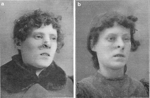
Fig. 2
Representative case from James H. Sequeiras report from 1903 showing a rodent ulcer on a 31-year-old woman before ( a ) and approximately 1 year after ( b ) X-ray therapy (reproduced from Br Med J, Sequeira J.H., Further observations upon the treatment of rodent ulcer by the X rays, vol. 2214, pp. 13071310, 1903 with permission from BMJ Publishing Group Ltd.)
Although the initial cathode ray tubes were somewhat erratic and unreliable in regard to the quality and intensity of their beams, new innovations in technology allowed for greater control of X-ray delivery via cathode ray tubes. In 1913 Coolidge introduced a modification of the cathode ray tube by increasing the vacuum and using a tungsten anode. These improvements allowed for a more reliable machine that could operate at higher voltages (150 kV) for longer periods of time. This led to the eventual development of a 200 kV machine in 1922 which enabled physicians to treat deeper tumors []. With the advent of these more reliable tubes, dermatologists such as George Miller MacKee served as pioneers in the field of radiation therapy for skin cancers and provided a benchmark textbook in 1921 entitled X-Rays and Radium in the Treatment of Skin Disease which, along with the subsequent editions, proved to be the gold standard for decades to come.
Before the discovery and widespread use of systemic and topical steroids, superficial radiation and Grenz ray therapy were both successfully utilized by dermatologists and non-dermatologists alike in the treatment of several benign yet recalcitrant skin conditions such as chronic inflammatory diseases, acne, and hirsutism [). This prompted the American Medical Associations Bureau of Investigation to get involved via investigations and public warnings in the 1930s. George Miller MacKees quote in the preface of his 1921 first edition textbook entitled X-Rays and Radium in the Treatment of Skin Disease proved to be a prescient charge for dermatologists and the medical community: Unfortunately they [x rays and radium] are dangerous agents in unskilled hands. Every physician who employs x rays and radium should have a thorough training and should possess modern knowledge and equipment.


