Background
Skin cancers are very common on the ear, due to its unprotected position on the body during outdoor activity, and continuous exposure to the sun through the car window while driving. The incidence of squamous cell carcinomas on the ear appears to be higher than that of basal cell carcinomas with reports suggesting squamous cell carcinomas being the most common (>50 %), followed by basal cell carcinomas (3040 %), and less frequently, melanomas(<5 %) [].
Many techniques have been discussed for reconstruction of the ear using flaps and grafts, after removal of skin cancer []. I am presenting a case of a malignant melanoma of the ear that needed a wider excision after excision of an initial 2 cm lesion (which was closed primarily with a wedge-excision after undermining) in this case a mastoid interpolation flap was used after the wider excision. The technique of the retro-auricular mastoid interpolation flap, its planning and useful tips are detailed in this article.
Case History
A 62-year old white female patient presented with a changing pigmented lesion on her R ear. Clinical examination and dermatoscopy suggested a probable malignant melanoma in situ and the lesion was excised. The histological examination revealed a melanoma-in-situ and a malignant melanoma Stage 1 A, Breslow thickness 0.3 mm, Clark level 2, non-ulcerated malignant melanoma.
Histopathology report:
EXCISION RIGHT EAR
Gross Description:
The specimen consists of a skin ellipse 24 x 14 x 5 mm with a central
variegated light and dark brown patch 15 x 6 mm.The entire lesion is processed.
SYNOPTIC REPORT FOR INVASIVE MALIGNANT MELANOMA
Tumour Type: Invasive malignant melanoma arising in an area of melanoma
in-situ
Clark Level: 2
Breslow Thickness: 0.3mm
Size of Invasive Tumour: 0.6mm width
Ulceration: Nil
Tumour Infiltrating Lymphocytes: Nil
Regression: Nil
Angiotropism: Nil
Lymphovascular Invasion: Nil
Perineural Spread/Neurotropism: Nil
Mitotic Rate: Not enough invasive tumour for a 1 sq mm count
Microscopic Satellitosis: Nil
Radial Margin of Excision: Margins clear of lesion.Closest melanoma
in-situ margin is 4mm.Closest invasive melanoma margin is 5mm.
Associated Nevus: Nil
SUMMARY DIAGNOSIS:
INVASIVE MALIGNANT MELANOMA, CLARK LEVEL 2, BRESLOW THICKNESS 0.375mm
MARGIN CLEAR
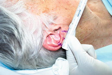
This tumor had an in-situ margin of 4 mm and the invasive melanoma had been removed with a margin of 5 mm. Margins for melanoma-in-situ have been the subject of recent debate. The accepted 5 mm guidelines were originally developed at a consensus meeting in 1992. A recent review in 2012, by a Mohs surgery team at a referral center for melanoma-in-situ suggested that the frequently recommended 5-mm margin for melanoma is inadequate. Standard surgical excision of melanoma in situ should include 9 mm of normal-appearing skin, similar to that recommended for early invasive melanoma [) avoiding the hair-bearing area. Given the original lesion was already 2 cm in diameter, the ear was already tissue deficient and this created an additional challenge.
Figure 1.1
Ear wide excision plan
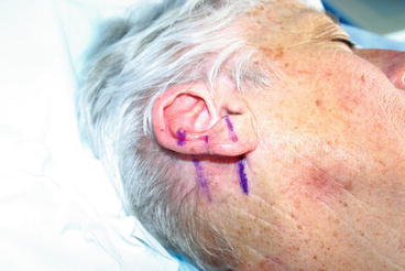
Figure 1.2
Ear interpolation flap plan
The Technique
The mastoid interpolation flap, which is the staged pedicle flap described herein, is very useful for helical ear defects when cartilage needs to be removed. It helps re-create a normal-looking ear. For smaller helical rim defects, a skin graft, cutaneous helical rim advancement flap, primary closure, or wedge resection often provides an excellent reconstructive result. Helical rim area can be a problem in itself with thin skin. However when cartilage needs to be removed, this increases the risk of perichondritis, which can be a surgical nuisance.
In a large series of patients after ear reconstruction, it was shown 24 % of cases become infected and may progress to perichondritis, if untreated [].
It is important to secure hemostasis during the procedure and place a drain to prevent hematoma formation. If perichondritis develops in spite of antibiotics, which is rare, then it is important to aggressively drain any abscess early. Fortunately, this is very rare after elective surgery for skin cancer.
As a general rule, the initial reconstructive effort is aimed only at repair of the anterior portion of the primary defect. Re-creation of the helical rim and posterior primary defect coverage is done at the second stage when the pedicle is detached and the ear is reconstructed. Some surgeons cut a template of foil or paper and lay over the mastoid to mark the outlines of the flap. It is important to avoid hair-bearing areas to avoid a hairy ear post-operatively. Rather than cutting out a template, I prefer to press the ear and lay it flat against the mastoid. Given that the excision margins are already marked on the ear, this allows to accurately plan the flap by continuing the markings onto the mastoid skin surface (Fig. ].
Figure 1.3
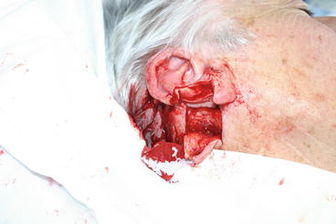
Ear mastoid interpolation flap being raised
Even with such sutures in place, it is often necessary to later on thin the flap to achieve perfect contours. I normally wait for 6 months post-operatively before planning any tertiary procedure such as this.
If the defect extends only to the helical rim, the flap can be started at the junction where mastoid skin meets the posterior ear. If the defect extends further medial to the scaphoid fossa or beyond, the flap incision is then ideally started on the posterior ear and extended onto the mastoid area. The flap should be sized slightly larger than the measured width of the defect and be long enough so that excessive tension is not placed on the flap after it is sutured [].
Once the flap is sutured in place, a drain is inserted to avoid any post-operative collection or hematoma formation. I tend to use a Penrose or a glove drain fashioned using a sterile surgical glove. Some authors prefer to use nasal packing or gel foam but in my experience, I have found this unnecessary. It is standard to apply a pressure dressing for 48 h, after which I usually remove the drain. I avoid suction drains (and prefer the glove drain) as the former are more bulky. I prefer to use a light dressing post-operatively which is not as noticeable (Fig. ).


