Eggert Stockfleth , Theodore Rosen and Stephen Shumack (eds.) Managing Skin Cancer 10.1007/978-3-540-79347-2_1 Springer-Verlag Berlin Heidelberg 2010
1. Diagnosis of Skin Cancer
S Astner 1
(1)
Department of Dermatology, Venerology and Allergology, CharitUniversty Hospital Berlin, Skin Cancer Center Charit, Charitplatz 1, Berlin, 10117, Germany
S Astner (Corresponding author)
Email:
1.1
1.1.1
1.1.2
1.1.3
1.1.4
1.2
1.2.1
1.2.2
1.2.3
1.2.4
1.3
1.3.1
1.3.2
1.3.3
1.3.4
Core Messages
Skin cancer diagnosis is based on clinical evaluation and histological exam of biopsy specimens represents the current diagnostic gold-standard. Recently non-invasive imaging techniques have emerged which may aid in the diagnosis and differential diagnosis of skin cancer.
Dermoscopy has been well established for the evaluation of pigmented lesions. As a novel diagnostic tool, reflectance confocal microscopy (RCM) has been applied for diagnosis of skin cancer and allows the evaluation of the skin at near histological resolution.
1.1 Clinical Evaluation and Risk Assessment
Diagnosis of cutaneous malignancies is based on the clinical evaluation of the patient, a detailed history, and ultimately, histological analysis. As the majority of epithelial skin tumors may already have been identi-fied by clinical evaluation, the total body skin exam is of utmost importance and should forego any invasive procedures. Lesion type, shape, demarcation, color, arrangement, and distribution should be recorded, with particular attention to aspects of asymmetry with respect to color and shape in pigmented lesions. Complete evaluations should include the palms and soles, the genital area, the scalp, and the lymph nodes.
A detailed history allows the assessment of the individual's skin cancer risk with regard to carcinogen exposure and familial cancer syndromes or risk factors. It should include a record of occupational and recreational sun exposure, a history of sunburns, the general health status, and a personal and family history of cancer. Congenital nevi, familial atypical moles, actinic damage, and Fitzpatrick skin type should be documented in each patient. By considering all aspects of history and clinical exam, patients at risk can be reliably identified for regular screening and prevention (Table ).
Table 1.1
Assessment of predisposing risk factors of skin cancer development
Risk factor | Differentiating criteria | Skin cancer risk | Recommendation |
|---|
Fitzpatrick skin phototype IIII/IV | Based on the ability to tan/burn none-(minimal-moderate-profuse) | SCC > BCC > MM | Primary/secondary prevention |
Screening |
Sunbathing habits | Use of sunscreen | SCC > BCC > MM | Education |
Summer vs. winter | Prevention |
Vacational vs occupational | Screening |
Use of tanning beds |
Area of residence | Latitude (proximally to equator), Altitude, urban vs industrial, Climatic conditions | SCC > BCC > MM | Education |
Prevention |
Screening |
Personal and family history of skin cancer | Ask for: NMSC, MM, NBCCS | MM, NMSC | Prevention |
Screening |
Common moles | Risk for melanoma increases with increasing number of moles (>100) | MM | Prevention |
Screening |
Atypical moles | Risk for melanoma; >5 or more atypical moles; familial dysplastic moles | MM | Monitoring |
Screening |
Changing mole | History | MM | Monitoring |
Screening |
Congenital moles | History | MM | Excision |
Gender of the patient | Male > female | MM, NMSC | Prevention |
Screening |
Specific risk factors | Arsenic, tar, radiotherapy, Use of photosensitizers, PUVA therapy or other phototherapies. | NMSC | Prevention |
Screening |
Other risk factors | History of smoking | NMSC | Screening |
Chronic/longstanding immunosuppression | MM? |
Nevus sebaceous | Clinical appearance of yellow, verrucous growth on the scalp, present since birth. | BCC | Excision |
Genetic syndromes with increased skin cancer risk | Epidermodysplasia verruciformis (EV) | HPV-related SCC AK, SCC, MM BCC MM AK, SCC | Close monitoring |
Xeroderma pigmentosum (XP) | Genetic counseling |
GorlinGoltz Syndrome (NBCCS) |
Familial multiple mole and melanoma (FAMM) |
Epidermolysis bullosa (dystrophic/junctional variants) |
An outline of risk factors to be assessed in a detailed evaluation of patients presenting for skin cancer screening and its practical implications
NMSC Nonmelanoma skin cancer; NBCCS nevoid basal cell carcinoma syndrome; HPV human papilloma virus; AK actinic kerato-sis; SCC squamous cell carcinoma; BCC basal cell carcinoma; MM malignant melanoma
1.1.1 Actinic Keratosis and Squamous Cell Carcinoma
Actinic keratoses (AK) present as multiple, erythema-tous to brown papules and plaques with adherent hyperkeratotic scales. In patients with extensive sun-exposure, entire fields of AK can be identified (Fig. ). Actinic cheilitis of the lower lip presents in the form of dry, fissured lips with marked atrophy, which may disrupt the vermillion border. Actinic cheilitis should be differentiated from allergic/toxic eczema of the lip, granulomatous cheilitis, erosive lichen planus, facti-cial processes, and ultimately, squamous cell carcinoma (SCC), on the basis of the history and clinical appearance.
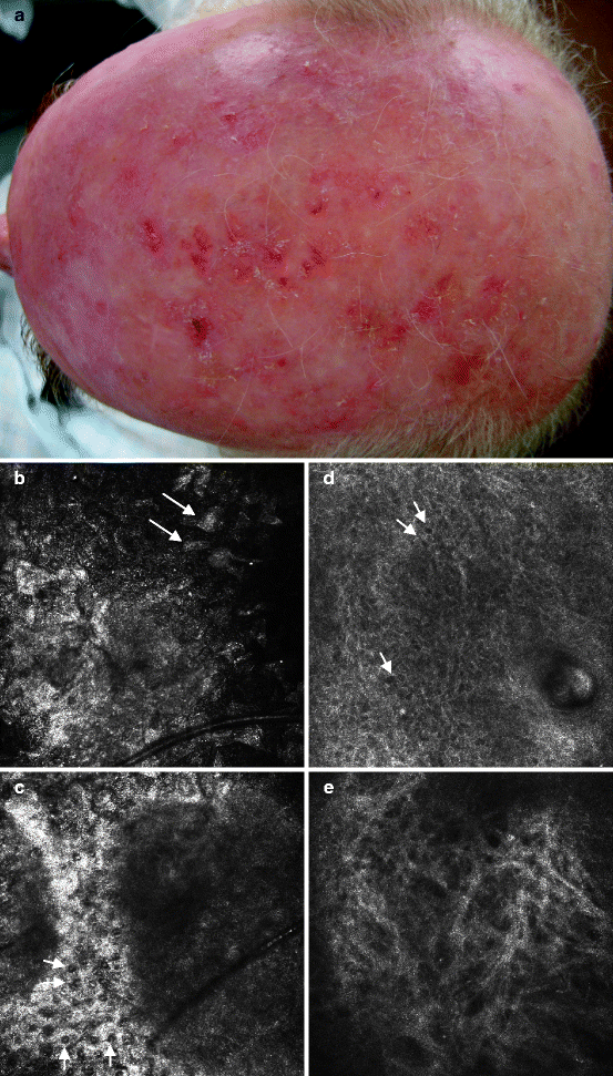
Fig. 1.1
( a ) Corresponds to clinical image of 67-year-old male, with a long standing history of sun-exposure. Clinical evaluation reveals numerous and confluent hyperkeratotic papules and plaques on sun-exposed areas of the balding scalp, consistent with actinic field cancerization. Normal skin is difficult to distinguish from clinically affected skin sites. Histology confirmed the diagnosis of actinic keratoses (AK). ( be ) Correspond to representative RCM images of AK. ( b ) RCM image obtained at the level of the stratum corneum with noted scaling and demarcation of individual corneocytes, seen as detached bright, polygonal structures ( white arrows ). ( c ) RCM image illustrating parakeratosis, identified by the dark nucleus placed centrally within the bright appearing corneocytes. The irregularities of the epidermal architecture are seen as dark hollow between bright rims of keratinocytes. ( d ) RCM image illustrating keratino-cyte pleomorphism, with atypical nuclei seen as dark round to oval, to polygonal structures ( white arrows ) of variable size and orientation. ( e ) RCM image obtained at the level of the mid-to-upper dermal layer illustrating solar elastosis. Irregular bundles of bright appearance and somewhat haphazard distribution correspond to irregular dermal elastic fibers. (RCM images obtained by VivaScope 1500, image dimensions 500500 m)

