• Complain

Jennifer T. Huang - Skin Tumors and Reactions to Cancer Therapy in Children

Here you can read online Jennifer T. Huang - Skin Tumors and Reactions to Cancer Therapy in Children full text of the book (entire story) in english for free. Download pdf and epub, get meaning, cover and reviews about this ebook. year: 2017, publisher: Springer, genre: Home and family. Description of the work, (preface) as well as reviews are available. Best literature library LitArk.com created for fans of good reading and offers a wide selection of genres:

Romance novel Science fiction Adventure Detective Science History Home and family Prose Art Politics Computer Non-fiction Religion Business Children Humor

Choose a favorite category and find really read worthwhile books. Enjoy immersion in the world of imagination, feel the emotions of the characters or learn something new for yourself, make an fascinating discovery.

Jennifer T. Huang Skin Tumors and Reactions to Cancer Therapy in Children

Skin Tumors and Reactions to Cancer Therapy in Children: summary, description and annotation

We offer to read an annotation, description, summary or preface (depends on what the author of the book "Skin Tumors and Reactions to Cancer Therapy in Children" wrote himself). If you haven't found the necessary information about the book — write in the comments, we will try to find it.

In todays era of genomic testing, targeted cancer therapies, and increased cancer survivorship, recognition and management of oncologic disease in children is becoming more important to address. This book familiarizes clinicians with the range of oncologic issues that pediatric dermatologists encounter, including the diagnosis and management of various skin tumors, cutaneous reactions to cancer therapies, and both acute and long term complications of cancer therapy. Written and edited by experts in the field, Pediatric Skin Tumors and Cutaneous Reactions to Cancer Therapy explores these important topics, beginning with the discussion of various skin tumors, and associated genetic syndromes, followed by chapters on acute cutaneous reactions to traditional and targeted cancer therapies, graft versus host disease, and opportunistic skin infections. This book concludes with the discussion of both malignant and nonmalignant late effects of the skin in childhood cancer survivors.

Jennifer T. Huang: author's other books


Who wrote Skin Tumors and Reactions to Cancer Therapy in Children? Find out the surname, the name of the author of the book and a list of all author's works by series.

Skin Tumors and Reactions to Cancer Therapy in Children — read online for free the complete book (whole text) full work

Below is the text of the book, divided by pages. System saving the place of the last page read, allows you to conveniently read the book "Skin Tumors and Reactions to Cancer Therapy in Children" online for free, without having to search again every time where you left off. Put a bookmark, and you can go to the page where you finished reading at any time.

Light

Font size:

Reset

Interval:

Bookmark:

Make
Springer International Publishing AG 2018
Jennifer T. Huang and Carrie C. Coughlin (eds.) Skin Tumors and Reactions to Cancer Therapy in Children
1. Melanoma and Spitz Nevi in Children
Catherine Warner 1
(1)
Department of Dermatology, Hahnemann Hospital, 219 N Broad Street, 4th Floor, Philadelphia, PA 19102, USA
(2)
Department of Pediatrics and Dermatology, Perelman School of Medicine at the University of Pennsylvania, Childrens Hospital of Philadelphia, 3401 Civic Center Boulevard, Room 3306, Philadelphia, PA 19104, USA
Catherine Warner
Email:
Melinda Jen (Corresponding author)
Email:
Keywords
Spitz nevus Atypical Spitz tumor Spitzoid tumor of unknown malignant potential Pediatric melanoma Fluorescence in situ hybridization Comparative genomic hybridization BAP1 HRAS BRAF Kinase fusion protein Kamino body CDKN2A TERT
Melanocytic nevi are common skin neoplasms and can be either congenital or acquired. While congenital nevi can be present at birth or appear shortly thereafter, acquired nevi continue to develop through childhood and into early adulthood. In a large cross-sectional study of kindergarten children in Germany, nevi were noted to increase in number from a median of 3 at 2 years of age to 19 at 7 years of age []. Since these two groups have similar skin types, this highlights the role of sun exposure in the development of nevi.
In addition to the development of new nevi, existing nevi naturally evolve throughout childhood, and a change in appearance may be not be concerning for melanoma []. Although pediatric melanoma is uncommon, identifying clinically suspicious lesions is essential for timely diagnosis and treatment. Appropriately identifying concerning lesions is especially important in the pediatric population where biopsies may be relatively more emotionally or psychologically stressful. Spitz nevi and atypical Spitz nevi pose a particular challenge clinically and histologically because of features that overlap with melanoma. Research has focused on histologic, immunohistochemical, and genomic methods to differentiate between these three entities.
Spitz Nevi
Key Points
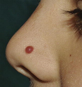
  • Most commonly found in the pediatric population.
  • Present as smooth pink, red, brown, or black papules that initially grow rapidly and then stabilize.
  • Composed of epithelioid and/or spindle cells.
In 1948, Dr. Sophie Spitz described a subset of pediatric melanocytic tumors that had features of melanoma but lacked their aggressive clinical characteristics []. Although previously called benign juvenile melanoma , this tumor is now called spindle and epithelioid cell nevus or, more commonly, Spitz nevus. A variant of the Spitz nevus is the pigmented spindle cell nevus (pigmented cell tumor of Reed, Reed nevus, pigmented Spitz nevus).
Clinical
Spitz nevi most commonly arise in patients less than 20 years of age [].
Fig 11 Characteristic appearance of a Spitz nevus as a smooth dome-shaped - photo 1
Fig. 1.1
Characteristic appearance of a Spitz nevus as a smooth dome-shaped pink papule []. Reprinted from Journal of the American Academy of Dermatology, Vol. 65, No. 6, Luo S, Sepehr A, Tsao H, Spitz nevi and other Spitzoid lesions: Part I. Background and Diagnoses, pages 10731084, 2011, with permission from Elsevier
On dermoscopy, Spitz nevi have several common patterns, including starburst, globular, homogeneous, negative network, and reticular patterns (Fig. ]. Common melanoma-specific structures, such as atypical pigment network, negative network, crystalline structures, blue-white veil, and irregular vessels, have been described in Spitz nevi but are more likely to be symmetric and organized.
Fig 12 Common dermoscopy patterns seen in Spitz nevi Reprinted from - photo 2
Fig. 1.2
Common dermoscopy patterns seen in Spitz nevi []. Reprinted from Dermatologic Clinics, Vol. 31, No. 2, Kerner M, Jaimes N, Scope A, Marghoob AA, Spitz nevi: a bridge between dermoscopic morphology and histopathology, pages 327335, 2013, with permission from Elsevier
Histology
While the majority of Spitz nevi are compound, they can also be junctional or intradermal. At low power, Spitz nevi are dome or wedge shaped, symmetric, and well-circumscribed. At higher power, Spitz nevi have characteristic epithelioid and/or spindled melanocytes with prominent cytoplasm. Junctional cells are nested and often have clefts separating individual cells or nests from adjacent epidermis. Cells mature to become small individual melanocytes deeper in the dermis. Kamino bodies are acellular pale eosinophilic structures often found within the epidermis and are composed of basement membrane material. Epidermal hyperplasia, hyperkeratosis, and hypergranulosis are commonly seen. Occasional mitotic figures can be seen within the epidermis or superficial dermis, although deep mitoses can indicate a more atypical tumor ( Fig. ) . Pigmented spindle cell nevi resemble Spitz nevi histologically but have prominent cytoplasmic pigmentation.
Fig 13 Spitz nevus a At low power the characteristic wedge-shaped - photo 3
Fig. 1.3
Spitz nevus. ( a ) At low power, the characteristic wedge-shaped, symmetric, well-circumscribed architecture of a Spitz nevus is seen (H&E, 40). ( b ) Junctional and dermal nests of mostly epithelioid and some spindled melanocytes are seen. Eosinophilic globules ( Kamino bodies) are in the epidermis, and there is mild epidermal hyperplasia. Lymphohistiocytic inflammation is present in the dermis (H&E, 100) (Image courtesy of Adam I. Rubin, MD)
Genetics
Approximately 20% of Spitz nevi demonstrate an increase in chromosome 11p, which is the site of the HRAS gene []. Gain of this genetic locus is very rare in cutaneous melanoma. Gain of chromosome 11p therefore suggests a more benign etiology (see Genetics and Molecular Analysis below). Apart from an occasional gain in HRAS , genetic gains or losses are rare in Spitz nevi.
Management
Although generally considered to be benign, controversy exists surrounding the management of Spitz nevi. For classic appearing Spitz nevi in school-aged children, some advocate for clinical observation, while others advocate a biopsy of any Spitzoid lesion []. Though, if re-excision is pursued, narrow 12 mm margins are adequate.
Atypical Spitz Tumors
Key Points
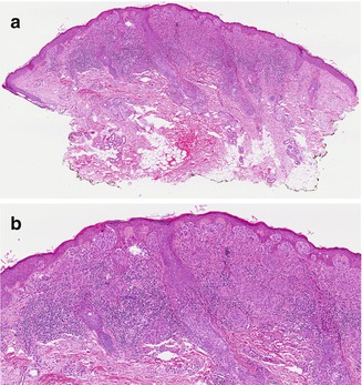
  • Histologic features overlapping with Spitz nevi and melanoma.
  • Molecular testing with fluorescence in situ hybridization and/or comparative genomic hybridization is an adjunct to histologic evaluation.
  • HRAS mutations, BAP1 mutations , and kinase fusions have been identified in these tumors.
Atypical Spitz tumors (AST), also called Spitzoid tumors of uncertain malignant potential (STUMP) , are challenging because they have histologic features that overlap with both classic Spitz nevi and with melanoma. Consequently, the diagnosis and management of these tumors are controversial and still evolving.
Atypical Spitz tumors are often considered within the larger category of borderline melanocytic tumors or melanocytic tumors of unknown malignant potential (MelTUMP). The term MelTUMP was first used by Elder and Xu to encompass a wide spectrum of lesions that demonstrate features of both benign melanocytic nevi and melanoma []. In addition to AST, pigmented epithelioid melanocytoma, deep penetrating nevus, and cellular blue nevus are the most common neoplasms included in this group.
Next page
Light

Font size:

Reset

Interval:

Bookmark:

Make

Similar books «Skin Tumors and Reactions to Cancer Therapy in Children»

Look at similar books to Skin Tumors and Reactions to Cancer Therapy in Children. We have selected literature similar in name and meaning in the hope of providing readers with more options to find new, interesting, not yet read works.


Reviews about «Skin Tumors and Reactions to Cancer Therapy in Children»

Discussion, reviews of the book Skin Tumors and Reactions to Cancer Therapy in Children and just readers' own opinions. Leave your comments, write what you think about the work, its meaning or the main characters. Specify what exactly you liked and what you didn't like, and why you think so.