Springer International Publishing Switzerland 2016
Michael T. Yen (ed.) Vascular Lesions of the Orbit and Face 10.1007/978-3-319-29704-0_1
1. Classification of Orbital Vascular Anomalies
Preeti J. Thyparampil 1 and Michael T. Yen 1
(1)
Department of Ophthalmology, Baylor College of Medicine, Houston, TX, USA
1.1 Introduction
Vascular lesions of the orbit comprise a significant portion of orbital pathology, accounting for 7 % in one review of 4000 orbital lesions []. A straightforward and functional classification of orbital vascular anomalies is important to aid clinicians in diagnosing these lesions and in planning treatment.
Current classification systems organize vascular lesions of the orbit based on histopathological features (tumors vs. malformations) and based on hemodynamic properties (no-flow, low-flow, or high-flow lesions). Additionally, current systems characterize vascular anomalies based on anatomic location or depth and complexity. While these various classification systems aid greatly in understanding vascular orbital anomalies, the nomenclature of some lesions remains confusing. For example, the cavernous hemangioma is better characterized as a venous flow (or low-flow) malformation as opposed to a hemangioma or tumor [].
While existing organizational systems do not eliminate all confusion in the classification of orbital vascular lesions, they nonetheless are very helpful for categorizing lesions and guiding treatment based on histopathological origin, clinical presentation, and radiographic imaging.
1.2 Tumors/Neoplasms vs. Malformations
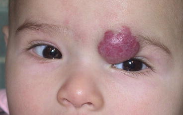
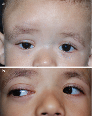
One classification system distinguishes vascular lesions as either tumors or malformations. Orbital vascular tumors, the most common of which is the infantile hemangioma (Fig..
Fig. 1.1
Large infantile hemangioma of the left orbit and upper eyelid
Table 1.1
Tumors vs. malformations
Tumors | Malformations |
|---|
Not present from birth | Present from birth |
Can grow in vitro | Cannot grow in vitro |
Proliferating endothelium | Dysplastic vessels without proliferating endothelium |
Well-circumscribed lobular masses on imaging | Less well circumscribed on imaging |
Infiltrate adjacent structures or cause mass effect on imaging | Distortion of adjacent structures on imaging by becoming incorporated into the vasculature of the nearby bone and soft tissue |
1.3 Classification of Malformations Based on Hemodynamics
The Orbital Society further advises classification of malformations based on flow characteristics. In this classification system, malformations can be characterized as either no-flow lesions, low-flow (venous,) lesions, or high-flow (arterial) lesions [].
Fig. 1.2
( a ) CT scan demonstrating dilated superior ophthalmic vein ( arrow ) in the right orbit. ( b ) Axial view of the same CT scan showing the dilated superior ophthalmic vein ( arrow ) in the right orbit
Based on these physical and imaging features, malformations can be characterized into three types. Very low or no-flow lesions are primarily lymphatic lesions and include venous lymphatic malformations or lymphangiomas (Fig. ].
Fig. 1.3
( a ) Extensive lymphangioma of the right forehead, brow, and orbit. ( b ) With anterior orbital extension, the lymphangioma may appear as a collection of cystic lesions in the conjunctiva
Additionally, malformations can be classified based on anatomical location. Superficial lesions usually involve just the eyelid or conjunctiva. Deep lesions are typically orbital without external visible components. Combined lesions have both superficial and deep components. Finally, complex lesions can be multifocal including orbital, intracranial, and systemic components and can be seen with multisystemic syndromes [.
Table 1.2
Classification of malformations based on hemodynamics
Low or no flow (lymphatic malformations) | Low flow (venous malformations) | High flow (arterial malformations) |
|---|
Purely lymphatic Combined venous lymphatic Lymphatic dominant Nondistensible i.e., lymphangiomas Venous dominant Distensible | Large connections to normal venous circulation Small connections to normal venous circulation i.e., cavernous hemangiomas | Direct connection from arterial side to venous side i.e., arteriovenous malformations i.e., congenital arteriovenous fistulas |
1.4 Very Low or No-Flow (Lymphatic) Vascular Malformations
These lesions derive embryologically from the venous system and undergo differentiation into a primarily lymphatic lesion. The ISSVA classification divides lymphatic lesions into microcystic, macrocystic, and mixed lesions. These malformations can also be characterized as purely lymphatic malformations and combined venous lymphatic malformations. Some combined lesions are lymphoid dominant, while others are venous dominant. Venous dominant lesions tend to be distensible, while lymphoid dominant lesions are not distensible and are commonly referred to as lymphangiomas. The deeper orbital portions of these mixed lesions tend be more venous. Venous dominant lesions tend to present at an earlier age (average 6 years), while lymphatic dominant lesions tend to present later (average age 13 years). Histopathological features of these vascular malformations include lymphorrhages, hemorrhages, lymphatic vessels, dysplastic venous channels, and smooth muscle [].
Lymphatic malformations and venolymphatic malformations vary in clinical appearance based on anatomic location. Superficial lesions of the conjunctiva may appear as clear cystic structures filled with xanthochromic material. Under the skin, they may appear bluish in color. These superficial lesions can usually be removed by surgical excision. These are typically considered solid, microcystic lymphangiomas. Direct excision can usually be performed without significant bleeding, but the lesion margins can be difficult to determine. Deep lesions can present with sudden proptosis due to hemorrhage or sudden increase in size related to concurrent illness. Sudden bouts of hemorrhage are due to capillary tufts present within macrocystic walls in the lesion. This is common with lymphatic dominant venolymphatic malformations (lymphangiomas). Management of these lesions may be warranted if sudden hemorrhage causes orbital congestion and compressive optic neuropathy, for ocular exposure secondary to proptosis, or for disfiguring appearance. Interventional treatment methods include simple aspiration and surgical excision. Surgical excision is more difficult with combined lesions and may require concomitant carbon dioxide laser treatment, gluing with fibrin or cyanoacrylate, or circumferential panorbitotomy [].

