• Complain

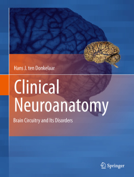
Hans J. Ten Donkelaar - Clinical Neuroanatomy: Brain Circuitry and Its Disorders

Here you can read online Hans J. Ten Donkelaar - Clinical Neuroanatomy: Brain Circuitry and Its Disorders full text of the book (entire story) in english for free. Download pdf and epub, get meaning, cover and reviews about this ebook. year: 2011, publisher: Springer Nature, genre: Home and family. Description of the work, (preface) as well as reviews are available. Best literature library LitArk.com created for fans of good reading and offers a wide selection of genres:

Romance novel Science fiction Adventure Detective Science History Home and family Prose Art Politics Computer Non-fiction Religion Business Children Humor

Choose a favorite category and find really read worthwhile books. Enjoy immersion in the world of imagination, feel the emotions of the characters or learn something new for yourself, make an fascinating discovery.

Hans J. Ten Donkelaar Clinical Neuroanatomy: Brain Circuitry and Its Disorders
  • Book:
    Clinical Neuroanatomy: Brain Circuitry and Its Disorders
  • Author:
  • Publisher:
    Springer Nature
  • Genre:
  • Year:
    2011
  • Rating:
    4 / 5
  • Favourites:
    Add to favourites
  • Your mark:
    • 80
    • 1
    • 2
    • 3
    • 4
    • 5

Clinical Neuroanatomy: Brain Circuitry and Its Disorders: summary, description and annotation

We offer to read an annotation, description, summary or preface (depends on what the author of the book "Clinical Neuroanatomy: Brain Circuitry and Its Disorders" wrote himself). If you haven't found the necessary information about the book — write in the comments, we will try to find it.

Connections define the functions of neurons: information flows along connections, as well as growth factors and viruses, and even neuronal death may progress through connections. Knowledge of how the various parts of the brain are interconnected to form functional systems is a prerequisite for the proper understanding of data from all fields in the neurosciences.

Clinical Neuroanatomy: Brain Circuitry and Its Disorders bridges the gap between neuroanatomy and clinical neurology. It emphasizes human and primate data in the context of disorders of brain circuitry which are so common in neurological practice. In addition, numerous clinical cases demonstrate how normal brain circuitry may be interrupted and to what effect. Following an introduction into the organization and vascularisation of the human brain and the techniques to study brain circuitry, the main neurofunctional systems are discussed, including the somatosensory, auditory, visual, motor, autonomic and limbic systems, the cerebral cortex and complex cerebral functions.

Hans J. Ten Donkelaar: author's other books


Who wrote Clinical Neuroanatomy: Brain Circuitry and Its Disorders? Find out the surname, the name of the author of the book and a list of all author's works by series.

Clinical Neuroanatomy: Brain Circuitry and Its Disorders — read online for free the complete book (whole text) full work

Below is the text of the book, divided by pages. System saving the place of the last page read, allows you to conveniently read the book "Clinical Neuroanatomy: Brain Circuitry and Its Disorders" online for free, without having to search again every time where you left off. Put a bookmark, and you can go to the page where you finished reading at any time.

Light

Font size:

Reset

Interval:

Bookmark:

Make
Hans J. ten Donkelaar Clinical Neuroanatomy Brain Circuitry and Its Disorders 10.1007/978-3-642-19134-3_1
Springer-Verlag Berlin Heidelberg 2011
1. Overview of the Human Brain and Spinal Cord
Hans J. ten Donkelaar 1
(1)
920 Department of Neurology/Clinical Neurophysiology, Radboud University Nijmegen Medical Centre, 9101, 6500 Nijmegen, The Netherlands
Hans J. ten Donkelaar
Email:
1.1
1.2
1.2.1
1.2.2
1.2.3
1.2.4
1.2.5
1.2.6
1.3
1.3.1
1.3.2
1.3.3
1.3.4
1.3.5
1.4
1.4.1
1.4.2
1.5
1.5.1
1.5.2
1.5.3
1.6
1.6.1
1.6.2
1.6.3
1.7
1.7.1
1.7.2
1.7.3
1.8
1.8.1
1.8.2
1.8.3
1.8.4
1.8.5
Abstract
During the last decades, there have been tremendous technical developments to study the human central nervous system (CNS) and its connectivity. Modern imaging techniques such as positron emission tomography (PET) and functional magnetic resonance imaging (fMRI) have greatly improved our knowledge of the circuitry of the human CNS. New developments in MR imaging such as diffusion tensor imaging (DTI) allow the visualization of at least the major fibre connections in the human CNS. Clinical Neuroanatomy tries to bridge the gap between neuroanatomy and clinical neurology and emphasizes human and primate data in the context of the many disorders of brain circuitry so common in neurological practice. In this introductory chapter, an overview is presented of the macroscopy (Sect. 1.2) and microscopy of the CNS and the relations of the brain to the skull, the skull base in particular (Sect. 1.3), and of the spinal cord to the vertebral column (Sect. 1.4). More in particular, the various structures of the brain and the spinal cord and their main fibre connections will be discussed, starting with the spinal cord (Sect. 1.4), followed by the brain stem (Sect. 1.5), the cerebellum (Sect. 1.6), the diencephalon (Sect. 1.7) and the telencephalon (Sect. 1.8).
1.1 Introduction
In this introductory chapter, an overview will be presented of the macroscopy and microscopy of the central nervous system (CNS). More in particular, the various structures of the brain and the spinal cord and their main fibre connections will be discussed. The vascularization of the brain and the spinal cord will be discussed in Chap. 2.
1.2 Macroscopy of the Brain
The lateral aspect of the brain is dominated by the cerebrum but also parts of the cerebellum and the brain stem are visible. On the medial aspect of the hemisected brain, the diencephalon becomes visible. The basal aspect of the brain lies on the base of the skull and is formed by the basal parts of the cerebrum, the brain stem and the cerebellum. The cerebrum consists of two cerebral hemispheres, each of which is composed of frontal, parietal, occipital and temporal lobes. The two hemispheres are separated by the interhemispheric fissure. Lateral, medial, dorsal and basal views of the brain will be discussed.
1.2.1 Lateral View of the Brain
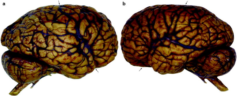
On the lateral aspect of the cerebrum (Fig. ).
Fig 11 Lateral views of the right a and left b hemispheres still - photo 1
Fig. 1.1
Lateral views of the right ( a ) and left ( b ) hemispheres, still enveloped by the leptomeninges. Note the difference between the slope of the lateral sulcus (indicated by lower arrow ). The upper arrow indicates the central sulcus (courtesy Pieter Wesseling and Arie Maat, Nijmegen)
In the frontal lobe , superior , middle and inferior frontal gyri , separated by superior and inferior frontal sulci, and the precentral gyrus are found (Fig. ; see Chap. 15).
The temporal lobe is composed of the superior , middle and inferior temporal gyri , separated by the superior and inferior temporal sulci (Fig. ; see Chap. 15). The triangular insula of Reil, described by Johann Christian Reil in 1809, lies in the depths of the lateral sulcus and is covered by the frontal, frontoparietal and temporal opercula. Its concave lower side is known as the limen insulae . The insula contains several vertically directed gyri, the gyri breves and longi .
On the lateral aspect of the parietal lobe , the postcentral gyrus , and the superior and inferior parietal lobules , separated by the intraparietal sulcus, are present. The inferior parietal lobule consists of the supramarginal and angular gyri (Fig..
1.2.2 Medial View of the Brain
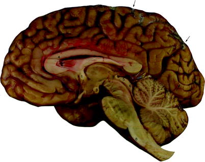
The prominent sulcus on the medial aspect of the cerebrum is the cingulate sulcus (the sulcus cinguli or scissure limbique of Broca ). Caudal to the paracentral lobule lies the large precuneus . The inner zone is formed by the cingulate gyrus , which is continuous through a narrowing (the isthmus gyri cinguli ) with the parahippocampal gyrus in the temporal lobe. The posterior part of the medial cerebral cortex has two deep sulci, which converge towards the splenium. The parieto - occipital sulcus separates the parietal and occipital lobes, and the calcarine sulcus divides the occipital lobe into a dorsal part (the cuneus ) and a ventral part (the lingual gyrus ). The primary visual cortex is mainly found above and below the calcarine sulcus.
Fig 12 Medial view of the right hemisphere after removal of the - photo 2
Fig. 1.2
Medial view of the right hemisphere after removal of the leptomeninges. The arrows indicate the central sulcus at the left and the parieto-occipital sulcus at the right. Indicated by numbers are: (1) anterior commissure; (2) posterior commissure; (3) mammillary body; (4) corpus callosum; (5) colliculus superior; (6) colliculus inferior; (7) pons; (8) pyramis (courtesy Pieter Wesseling and Arie Maat, Nijmegen)
In the midsagittally cut brain, the most prominent structure is the corpus callosum (Fig. ). Different parts of this structure are referred to as the rostrum , the genu , the truncus and the splenium . The most rostral part is the rostrum, which borders on the lamina terminalis. The truncus is the middle part, whereas the splenium forms the thick caudal part. Below the corpus callosum, the septum pellucidum and the fornix are found. The third ventricle shows three recesses, the infundibular recess into the upper part of the hypophysial stalk, the optic recess above the optic chiasm, and the pineal recess below the pineal gland (the epiphysis cerebri ). The third ventricle continues caudally via the aqueductus cerebri of the mesencephalon into the fourth ventricle . Above the cerebral aqueduct, the superior and inferior colliculi are found and, dorsal to the fourth ventricle, the cerebellum . The fourth ventricle intrudes into the cerebellum as the fastigium . Between the inferior colliculus and the cerebellum, the roof of the fourth ventricle is formed by a thin membrane, the velum medullare superius on which lies the lingula of the cerebellum. The caudal part of the roof of the fourth ventricle is formed by the tela choroidea and the plexus choroideus.
1.2.3 Dorsal View of the Brain
In the brain, viewed from above, the frontal, parietal and occipital lobes are visible (Fig. ). The cerebral hemispheres are separated by the longitudinal interhemispheric fissure . In the frontal and occipital regions, the separation is complete but centrally the fissure extends only to the broad interhemispheric commissure, the corpus callosum. Almost halfway between the frontal and occipital poles of each hemisphere, the central sulcus courses from the medial margin of the dorsal surface of the brain towards the lateral sulcus. Parallel to this sulcus are located the precentral gyrus rostrally and the postcentral gyrus caudally. These gyri are bordered by the precentral and postcentral sulci, respectively. In front of the precentral gyrus are, from medial to lateral, the superior frontal gyrus, the superior frontal sulcus, the middle frontal gyrus, the inferior frontal sulcus and the inferior frontal gyrus. Caudal to the postcentral sulcus can be distinguished the superior and inferior parietal lobules, the parieto-occipital sulcus and the occipital gyri.
Next page
Light

Font size:

Reset

Interval:

Bookmark:

Make

Similar books «Clinical Neuroanatomy: Brain Circuitry and Its Disorders»

Look at similar books to Clinical Neuroanatomy: Brain Circuitry and Its Disorders. We have selected literature similar in name and meaning in the hope of providing readers with more options to find new, interesting, not yet read works.


Reviews about «Clinical Neuroanatomy: Brain Circuitry and Its Disorders»

Discussion, reviews of the book Clinical Neuroanatomy: Brain Circuitry and Its Disorders and just readers' own opinions. Leave your comments, write what you think about the work, its meaning or the main characters. Specify what exactly you liked and what you didn't like, and why you think so.