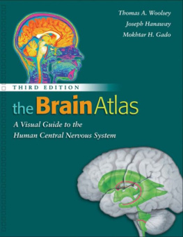
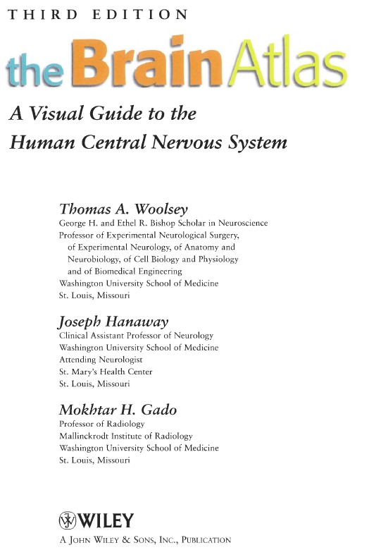
Thomas A. Woolsey - The Brain Atlas: A Visual Guide to the Human Central Nervous System
Here you can read online Thomas A. Woolsey - The Brain Atlas: A Visual Guide to the Human Central Nervous System full text of the book (entire story) in english for free. Download pdf and epub, get meaning, cover and reviews about this ebook. year: 2007, publisher: Wiley-Liss, genre: Home and family. Description of the work, (preface) as well as reviews are available. Best literature library LitArk.com created for fans of good reading and offers a wide selection of genres:
Romance novel
Science fiction
Adventure
Detective
Science
History
Home and family
Prose
Art
Politics
Computer
Non-fiction
Religion
Business
Children
Humor
Choose a favorite category and find really read worthwhile books. Enjoy immersion in the world of imagination, feel the emotions of the characters or learn something new for yourself, make an fascinating discovery.

- Book:The Brain Atlas: A Visual Guide to the Human Central Nervous System
- Author:
- Publisher:Wiley-Liss
- Genre:
- Year:2007
- Rating:5 / 5
- Favourites:Add to favourites
- Your mark:
The Brain Atlas: A Visual Guide to the Human Central Nervous System: summary, description and annotation
We offer to read an annotation, description, summary or preface (depends on what the author of the book "The Brain Atlas: A Visual Guide to the Human Central Nervous System" wrote himself). If you haven't found the necessary information about the book — write in the comments, we will try to find it.
The Brain Atlas: A Visual Guide to the Human Central Nervous System truly integrates modern neuroscience with clinical practice and is now completely revised and updated in its Third Edition. Now more than ever, it is the best available visual guide to human neuroanatomy for undergraduate and graduate medical students, clinicians and psychologists.
It flows logically from surface anatomy to cross-sections, and then on to regional histology, ending with diagrams of the major neuronal systems responsible for the brains magnificent array of functions. The books five sections cover Background Information, The Brain and Its Blood, Brain Slices, Histological Sections, and Pathways.
The Brain Atlas: A Visual Guide to the Human Central Nervous System, Third Edition features:
- A large number of new images reflecting the latest updates in imaging equipment and techniques
- Unrivalled treatment of brain pathways, including meticulous blood supply maps
- Systematic use of magnetic resonance images side-by-side with corresponding brain slices
- A direct labeling system, including an alphabetical list of terms for each image
From the reviews of the Second Edition:
an essential requirement for the library of any individual who works in the field... if you buy only one atlas, this is the one to buy. JOURNAL OF NEUROSURGERY
an excellent tool for understanding the central nervous system. It is a good companion at every level of training and for health care professionals. ARCHIVES OF NEUROLOGY
Thomas A. Woolsey: author's other books
Who wrote The Brain Atlas: A Visual Guide to the Human Central Nervous System? Find out the surname, the name of the author of the book and a list of all author's works by series.


The Brain Atlas: A Visual Guide to the Human Central Nervous System — read online for free the complete book (whole text) full work
Below is the text of the book, divided by pages. System saving the place of the last page read, allows you to conveniently read the book "The Brain Atlas: A Visual Guide to the Human Central Nervous System" online for free, without having to search again every time where you left off. Put a bookmark, and you can go to the page where you finished reading at any time.
Font size:
Interval:
Bookmark:
Copyright 2008 by John Wiley & Sons, Inc. All rights reserved.
Published by John Wiley & Sons, Inc., Hoboken, New Jersey.
Published simultaneously in Canada.
No part of this publication may be reproduced, stored in a retrieval system, or transmitted in any form or by any means, electronic, mechanical, photocopying, recording, scanning, or otherwise, except as permitted under Section 107 or 108 of the 1976 United States Copyright Act, without either the prior written permission of the Publisher, or authorization through payment of the appropriate per-copy fee to the Copyright Clearance Center, Inc., 222 Rosewood Drive, Danvers, MA 01923, 978-750-8400, fax 978-750-4470, or on the web at
Limit of Liability/Disclaimer of Warranty: While the publisher and author have used their best efforts in preparing this book, they make no representations or warranties with respect to the accuracy or completeness of the contents of this book and specifically disclaim any implied warranties of merchantability or fitness for a particular purpose. No warranty may be created or extended by sales representatives or written sales materials. The advice and strategies contained herein may not be suitable for your situation. You should consult with a professional where appropriate. Neither the publisher nor author shall be liable for any loss of profit or any other commercial damages, including but not limited to special, incidental, consequential, or other damages.
For general information on our other products and services please contact our Customer Care Department within the U.S. at 877-762-2974, outside the U.S. at 317-572-3993 of fax 317-572-4002.
Wiley also publishes its books in a variety of electronic formats. Some content that appears in print, however, may not be available in electronic format.
Library of Congress Cataloging-in-Publication Data:
Woolsey, Thomas A.
The brain atlas: a visual guide to the human central nervous system / Thomas A. Woolsey,
Joseph Hanaway, Mokhtar H. Gado. 3rd ed.
p. cm.
Previous ed. cataloged under title.
Includes bibliographical references and index.
ISBN 978-0-470-08476-2
1. Brain Anatomy Atlases. 2. Central nervous system Anatomy Atlases. I. Hanaway, Joseph,
1933II. Gado, Mokhtar H., 1931-III. Title.
QM455.B633 2003
611.00222 dc21
2002013579
Dedicated to
Clinton N. Woolsey
Jerzy E. Rose
David Bodian
W. Maxwell Cowan
Raymond D. Adams
C. Miller Fisher
James W. Bull
George du Boulay
Contents
Preface
The first edition of TheBrainAtlas:A VisualGuidetotheHumanCentralNervousSystem (1997) gave students of modern neuroscience an innovative combination of information on the human brain from many different sources. It included: dissections of the human brain and its blood vessels; photographs of human brain slices; MRI scans matched directly to those slices; maps of the brain territories supplied by major blood vessels; histological sections through the spinal cord, brain stem, thalamus, hypothalamus, and forebrain; and pathway diagrams rendered to convey the three-dimensional character of the principal connections of the brain. The first edition of TheBrainAtlas also was published in French and Italian. The second edition of TheBrainAtlas (2002) added several major improvements: simplified layout; a novel labeling system with direct labeling of all structures and all terms listed alphabetically on or opposite each page for rapid identification; reformatted pathway diagrams for improved clarity; and new illustrations to show cortical sulcal patterns and fiber bundles in the brain. Further, the second edition of TheBrainAtlas was spiral bound to lie flat to make it easier to use and more durable.
Many medical schools, universities, and colleges adopted the TheBrainAtlas for use in their neuroscience courses. The second edition was adopted by more institutions and is increasingly used as a resource in more undergraduate and graduate courses and health care training programs. Critical feedback from colleagues, instructors, medical, graduate, and undergraduate students, residents, and others has been given on both editions. These suggestions and the expertise of our publisher have guided the preparation of this third edition.
The third edition of TheBrainAtlas follows the successful organization, layout and design of the previous editions. However, there are a number of important improvements. First, newer imaging techniques have greatly improved in quality and detail, such that one is often hard pressed to distinguish radiological images from living human beings from those of brain slices prepared painstakingly from fixed brains. The third edition of The TheBrainAtlas has completely new radiological images of brain sections and blood vessels. Further, magnetic resonance images (MRI) have been added to illustrate the brain stem and spinal cord for comparison to the histological sections they match. Second, imaging is now fully integrated into research and clinical care so much so that the orientation conventions of radiology are now the standard. The third edition of TheBrainAtlas reflects this. The histological sections have been rotated 180 consistent with radiological conventions. In this orientation, axial images of the brain are conceptually continuous with the histological sections from the midbrain to the spinal cord. Accordingly, the order of the sections in Part III is now rearranged to flow directly from axial brain images into histological cross-sections through brain stem and spinal cord in Part IV. Third, this new edition of TheBrainAtlas includes several new and/or significantly revised illustrations in Part V that reflect advances in understanding the brain and its disorders. These are related to: a) the basal gangliasubstantia nigra, subthlamic nucleus, thalamus-cortical circuits; b) brain pathways for arousal and sleep; and c) pain pathways. Fourth, new diagrams integrating sections through the thalamus and hypothalamus illustrate the layout of cell groups and their connections in those structures. These are conceptually similar to the illustrations showing the organization of cranial nerve nuclei in the brain stem. Lastly, the third edition of TheBrainAtlas is printed so that complementary figures showing the organization of the brain stem cranial nerves and nuclei are on facing pages for direct comparison.
The third edition of TheBrainAtlas is designed to be an even easier to use, up-to-date learning resource for students of medicine and other health care professions (Physical Therapy, Occupational Therapy, Nursing), and students of biology, neuroscience, psychology and philosophy at all stages of their education. It is our hope that the third edition of TheBrainAtlas will continue to serve as an authoritative reference that is easy to use. This is important for anyone interested in the human brain, particularly resident and staff neuropathologists, neuroradiologists, neurologists, neurosurgeons and psychiatrists; selected other health care personnel; and neurobiologists, psychologists and cognitive scientists. Now is a remarkable epoch for the study of the human brain. It is our hope that the third edition of
Next pageFont size:
Interval:
Bookmark:
Similar books «The Brain Atlas: A Visual Guide to the Human Central Nervous System»
Look at similar books to The Brain Atlas: A Visual Guide to the Human Central Nervous System. We have selected literature similar in name and meaning in the hope of providing readers with more options to find new, interesting, not yet read works.
Discussion, reviews of the book The Brain Atlas: A Visual Guide to the Human Central Nervous System and just readers' own opinions. Leave your comments, write what you think about the work, its meaning or the main characters. Specify what exactly you liked and what you didn't like, and why you think so.