Skandalakis Lee John - Surgical anatomy and technique a pocket manual
Here you can read online Skandalakis Lee John - Surgical anatomy and technique a pocket manual full text of the book (entire story) in english for free. Download pdf and epub, get meaning, cover and reviews about this ebook. City: New York, year: 2009, publisher: Springer, genre: Home and family. Description of the work, (preface) as well as reviews are available. Best literature library LitArk.com created for fans of good reading and offers a wide selection of genres:
Romance novel
Science fiction
Adventure
Detective
Science
History
Home and family
Prose
Art
Politics
Computer
Non-fiction
Religion
Business
Children
Humor
Choose a favorite category and find really read worthwhile books. Enjoy immersion in the world of imagination, feel the emotions of the characters or learn something new for yourself, make an fascinating discovery.

- Book:Surgical anatomy and technique a pocket manual
- Author:
- Publisher:Springer
- Genre:
- Year:2009
- City:New York
- Rating:3 / 5
- Favourites:Add to favourites
- Your mark:
Surgical anatomy and technique a pocket manual: summary, description and annotation
We offer to read an annotation, description, summary or preface (depends on what the author of the book "Surgical anatomy and technique a pocket manual" wrote himself). If you haven't found the necessary information about the book — write in the comments, we will try to find it.
Skandalakis Lee John: author's other books
Who wrote Surgical anatomy and technique a pocket manual? Find out the surname, the name of the author of the book and a list of all author's works by series.


Surgical anatomy and technique a pocket manual — read online for free the complete book (whole text) full work
Below is the text of the book, divided by pages. System saving the place of the last page read, allows you to conveniently read the book "Surgical anatomy and technique a pocket manual" online for free, without having to search again every time where you left off. Put a bookmark, and you can go to the page where you finished reading at any time.
Font size:
Interval:
Bookmark:
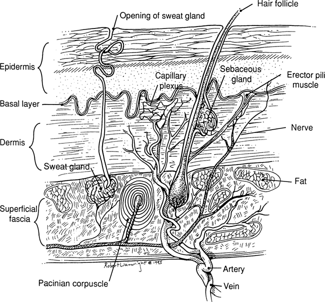
- The epidermis is avascular.
- The dermis is tough, strong, and very vascular.
- The superficial fascia is the subcutaneous tissue that blends with the reticular layer of the dermis.
- The principal blood vessels of the skin lie in subdermal areas.
- The basement membrane is the lowest layer of the epidermis.
- The papillary dermis is the upper (superficial) layer of the dermis, just below the basement membrane.
- The reticular dermis is the lower (deep) layer of the dermis, just above the fat.
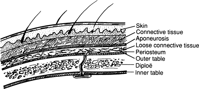
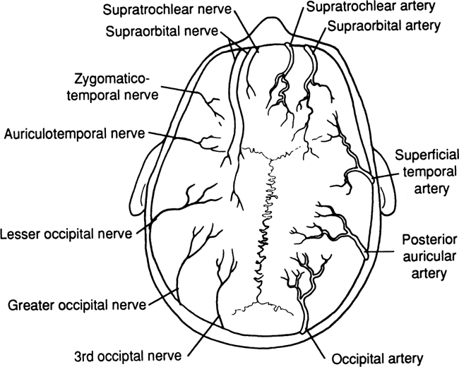
- The blood supply of the scalp is rich. Arteries are anastomosed very freely.
- The arteries and veins travel together in a longitudinal fashion.
- A transverse incision or laceration will produce a gap. Dangerous bleeding will take place from both vascular ends due to nonretraction of the arteries by the close, dense, connective layer.
- Always repair the aponeurotic galea to avoid hematoma under it.
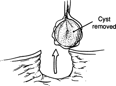
- With elective cases (excision of sebaceous cysts, etc.), whenever possible, make a longitudinal incision.
- Drain infections promptly. Use antibiotics to prevent intracranial infections via the emissary veins.
- Shave 12 cm around the site of the incision or laceration.
- After cleansing the partially avulsed scalp, replace it and dbride the wound; then suture with nonabsorbable sutures.
- Use pressure dressing as required. Sutures may be removed in 35 days.
- Be sure about the diagnosis. A very common sebaceous cyst could be an epidermoid cyst of the skull involving the outer or inner table, or both, with extension to the cerebral cortex. In such a case, call for a neurosurgeon. The best diagnostic procedure is an AP and lateral film of the skull to rule out bony involvement.
- Because the skin, connective tissue, and aponeurosis are so firmly interconnected, for practical purposes, they form one layer: the surgical zone of the scalp.
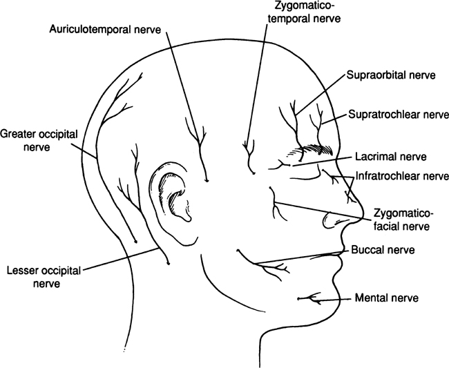
- Lesser occipital (second and third ventral nerves)
- Greater occipital (second and third dorsal nerves)
- Auriculotemporal (mandibular nerve)
- Zygomaticotemporal, zygomaticofacial (zygomatic [maxillary] nerve)
- Supraorbital (ophthalmic nerve)
- Supratrochlear (ophthalmic nerve)
Font size:
Interval:
Bookmark:
Similar books «Surgical anatomy and technique a pocket manual»
Look at similar books to Surgical anatomy and technique a pocket manual. We have selected literature similar in name and meaning in the hope of providing readers with more options to find new, interesting, not yet read works.
Discussion, reviews of the book Surgical anatomy and technique a pocket manual and just readers' own opinions. Leave your comments, write what you think about the work, its meaning or the main characters. Specify what exactly you liked and what you didn't like, and why you think so.