Smith - The tales teeth tell: development, evolution, behavior
Here you can read online Smith - The tales teeth tell: development, evolution, behavior full text of the book (entire story) in english for free. Download pdf and epub, get meaning, cover and reviews about this ebook. City: Cambridge;Massachusetts, year: 2018, publisher: MIT Press, genre: Home and family. Description of the work, (preface) as well as reviews are available. Best literature library LitArk.com created for fans of good reading and offers a wide selection of genres:
Romance novel
Science fiction
Adventure
Detective
Science
History
Home and family
Prose
Art
Politics
Computer
Non-fiction
Religion
Business
Children
Humor
Choose a favorite category and find really read worthwhile books. Enjoy immersion in the world of imagination, feel the emotions of the characters or learn something new for yourself, make an fascinating discovery.

- Book:The tales teeth tell: development, evolution, behavior
- Author:
- Publisher:MIT Press
- Genre:
- Year:2018
- City:Cambridge;Massachusetts
- Rating:4 / 5
- Favourites:Add to favourites
- Your mark:
The tales teeth tell: development, evolution, behavior: summary, description and annotation
We offer to read an annotation, description, summary or preface (depends on what the author of the book "The tales teeth tell: development, evolution, behavior" wrote himself). If you haven't found the necessary information about the book — write in the comments, we will try to find it.
Smith: author's other books
Who wrote The tales teeth tell: development, evolution, behavior? Find out the surname, the name of the author of the book and a list of all author's works by series.


The tales teeth tell: development, evolution, behavior — read online for free the complete book (whole text) full work
Below is the text of the book, divided by pages. System saving the place of the last page read, allows you to conveniently read the book "The tales teeth tell: development, evolution, behavior" online for free, without having to search again every time where you left off. Put a bookmark, and you can go to the page where you finished reading at any time.
Font size:
Interval:
Bookmark:
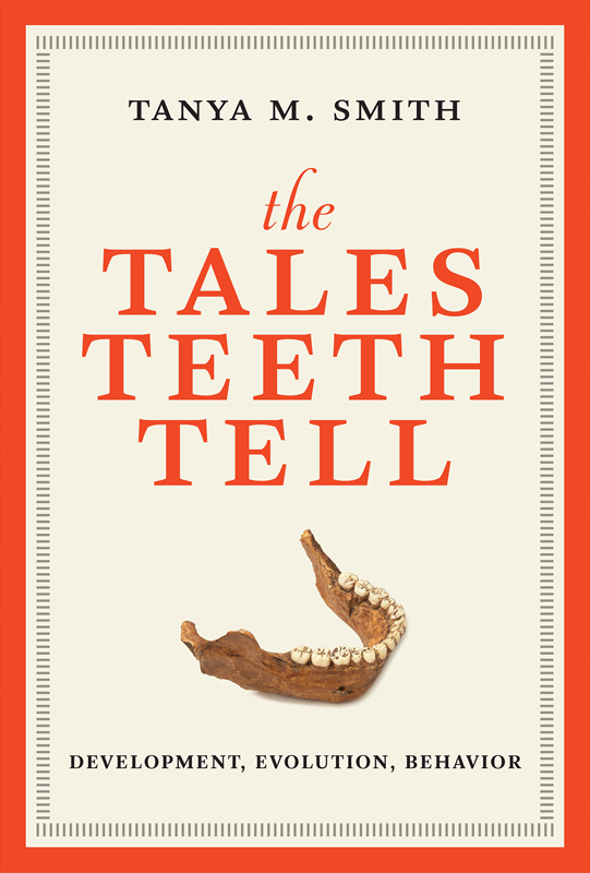
The Tales Teeth Tell
Development, Evolution, Behavior
The MIT Press
Cambridge, Massachusetts
London, England
2018 Massachusetts Institute of Technology
All rights reserved. No part of this book may be reproduced in any form by any electronic or mechanical means (including photocopying, recording, or information storage and retrieval) without permission in writing from the publisher.
This book was set in Stone Serif by Westchester Publishing Services. Printed and bound in the United States of America.
Library of Congress Cataloging-in-Publication Data
Names: Smith, Tanya M., author.
Title: The tales teeth tell : development, evolution, behavior / Tanya M. Smith.
Description: Cambridge, MA : The MIT Press, [2018] | Includes bibliographical references and index.
Identifiers: LCCN 2018005095 | ISBN 9780262038713 (hardcover : alk. paper)
Subjects: LCSH: Dental anthropology. | Dental calculus--Study and teaching. | Teeth--History.
Classification: LCC GN209 .S55 2018 | DDC 599.9/43--dc23 LC record available at https://lccn.loc.gov/2018005095
To my friend and mentor Donald Reid, for his steadfast support and inexhaustible love of teeth.
Contents
List of Figures
Growth lines inside a 9- to 12-million-year-old fossilized ape molar. Rhythms ranging from 9 days (broad diagonal lines) to 24 hours (small, light and dark, box-like features stacked vertically) reveal that this crown took more than 2 years to form. The colorization is due to the use of polarized light microscopy, highlighting variation in mineral structure. Fossil courtesy of the Natural History Museum (London).
Infant Neanderthal upper jaw and associated baby and permanent teeth. Individuals from this Belgian site (commonly known as Schmerling Cave or the second Engis cave) were the first fossil hominins ever discovered. Fossil courtesy of the University of Lige (Belgium).
Infant skull from the most complete australopithecine child discovered. Upper image is the original fossil; lower image is a virtual model showing its erupted baby teeth after sandstone removal. Fossil courtesy of the National Museum of Ethiopia (Addis Ababa). Image credit: Zeray Alemseged and Paul Tafforeau.
The Australopithecus africanus child studied by Raymond Dart. The upper and lower jaws have a full set of baby teeth along with the permanent first molars. Fossil courtesy of the University of the Witwatersrand (Johannesburg).
Enamel microstructure in a fossil tooth imaged with scanning electron microscopy. Faint vertical growth lines in the center of the image bisect diagonal enamel prisms. Fossil courtesy of Bob Anemone.
, below.
Classic illustration of the stages of tooth development and use. Progressive stages from left to right: cellular organization (labeled growth), enamel and dentine secretion (calcification), root formation (eruption), and wear (attrition). Black regions represent the mineralizing jawbone, which develops around the tooth as it is forming. Reproduced with permission from Isaac Schour and Maury Massler, Studies in Tooth Development: The Growth Pattern of Human Teeth, Part 1, Journal of the American Dental Association 27 (1940): 17781793.
Transmission electron micrograph of enamel-forming cells secreting enamel prisms. Enamel-forming cells run diagonally and secrete enamel prisms (EP) near the top of the image, which are made up of thin crystallites. Darkly stained round nuclei are visible on the opposite end of the cells. Modified from Erik Rnnholm, An Electron Microscopic Study of the Amelogenesis in Human Teeth: I. The Fine Structure of the Ameloblasts, Journal of Ultrastructure Research 6 (1962): 229248, with permission from Elsevier.
Magnified enamel showing the geometry of enamel prisms. (A) Scanning electron microscope image of chimpanzee enamel showing the three-dimensional nature of enamel prisms running from left to right. (B) Light micrograph of enamel prisms (slightly curved vertical lines) running from dentine (dark conical tip) to the enamel surface (upper boundary), a distance of more than 2.5 millimeters in this human molar. Reprinted from Paul Tafforeau, John P. Zermeno, and Tanya M. Smith, Tracking Cellular-Level Enamel Growth and Structure in 4D with Synchrotron Imaging, Journal of Human Evolution 62, no. 3 (2012): 424428, with permission from Elsevier. Chimpanzee specimen courtesy of Lawrence Martin.
Molar thin section showing the hard tissues: enamel (E), dentine (D), and cementum (C). Also included are the periodontal ligament (PDL) and surrounding bone (B). Lower-right inset shows vertical light and dark cementum annulations magnified with polarized light microscopy. Chimpanzee specimen courtesy of Donald Reid.
Experimental demonstration of daily lines in primate tooth enamel. A xylenol orange (XO) label was administered and then followed 8 days later by a minocycline (M) injection, and eight daily lines (red labels) appear between injections. Modified and reprinted from Tanya M. Smith, Experimental Determination of the Periodicity of Incremental Features in Enamel, Journal of Anatomy 208, Issue 1 (January 2006): 99113, with permission from Wiley-Liss. Macaque specimen courtesy of Joyce Sirianni.
Microscopic biological rhythms in primate enamel and dentine. Long-period lines inside teeth are indicated with white arrows in enamel (A) and dentine (B), and by blue dotted lines on the outsides of the tooth crown (C) and root (D). Four daily lines are indicated within each white bracket of A and B. Reprinted from Tanya M. Smith and Paul Tafforeau, New Visions of Dental Tissue Research, Evolutionary Anthropology 17 (2008): 213226, with permission from Wiley-Liss. Fossils courtesy of the Natural History Museum (London) and the University of Lige (Lige).
Molar before and after cutting to produce a section for microscopic imaging. Reprinted from Tanya M. Smith and Paul Tafforeau, New Visions of Dental Tissue Research, Evolutionary Anthropology 17 (2008): 213226, with permission from Wiley-Liss. Chimpanzee specimens courtesy of Christophe Boesch and Lawrence Martin.
prior to cutting. (A) 3D rendering of the chewing surface; (B) cross-sectional slice through the front cusps; (C) 3D model of the tooth showing the enamel cap (transparent yellow), enamel-dentine junction (red), and dentine (transparent blue); (D) isolated enamel cap; (E) isolated enamel-dentine junction; (F) isolated dentine under the enamel. Reprinted from Tanya M. Smith and Paul Tafforeau, New Visions of Dental Tissue Research, Evolutionary Anthropology 17 (2008): 213226, with permission from Wiley-Liss. Chimpanzee specimen courtesy of Christophe Boesch.
.) Enamel prisms run from the dentine (lower right) to the tooth surface (upper left). Individual continuous prisms are represented by unique colors, which have been traced with software. Daily lines can be seen as fine, paired light and dark bands in the left side of the background. Modified from Paul Tafforeau, John P. Zermeno, and Tanya M. Smith, Tracking Cellular-level Enamel Growth and Structure in 4D with Synchrotron Imaging, Journal of Human Evolution 62, no. 3 (2012): 424428, with permission from Elsevier. Chimpanzee specimen courtesy of Lawrence Martin.
Naturally shed baby tooth showing the neonatal (birth) line. The upper image shows the enamel crown and remaining root dentine. A small white box reveals the area imaged at higher magnification in the panel below, where the neonatal line (white arrow) runs toward the worn surface of the tooth. Human tooth courtesy of Charis Ng.
Font size:
Interval:
Bookmark:
Similar books «The tales teeth tell: development, evolution, behavior»
Look at similar books to The tales teeth tell: development, evolution, behavior. We have selected literature similar in name and meaning in the hope of providing readers with more options to find new, interesting, not yet read works.
Discussion, reviews of the book The tales teeth tell: development, evolution, behavior and just readers' own opinions. Leave your comments, write what you think about the work, its meaning or the main characters. Specify what exactly you liked and what you didn't like, and why you think so.