David Roberts MSPAS RN PA-C - Mastering the 12-Lead EKG, Second Edition
Here you can read online David Roberts MSPAS RN PA-C - Mastering the 12-Lead EKG, Second Edition full text of the book (entire story) in english for free. Download pdf and epub, get meaning, cover and reviews about this ebook. year: 2019, publisher: Springer Publishing Company, Inc., genre: Home and family. Description of the work, (preface) as well as reviews are available. Best literature library LitArk.com created for fans of good reading and offers a wide selection of genres:
Romance novel
Science fiction
Adventure
Detective
Science
History
Home and family
Prose
Art
Politics
Computer
Non-fiction
Religion
Business
Children
Humor
Choose a favorite category and find really read worthwhile books. Enjoy immersion in the world of imagination, feel the emotions of the characters or learn something new for yourself, make an fascinating discovery.

- Book:Mastering the 12-Lead EKG, Second Edition
- Author:
- Publisher:Springer Publishing Company, Inc.
- Genre:
- Year:2019
- Rating:5 / 5
- Favourites:Add to favourites
- Your mark:
Mastering the 12-Lead EKG, Second Edition: summary, description and annotation
We offer to read an annotation, description, summary or preface (depends on what the author of the book "Mastering the 12-Lead EKG, Second Edition" wrote himself). If you haven't found the necessary information about the book — write in the comments, we will try to find it.
David Roberts MSPAS RN PA-C: author's other books
Who wrote Mastering the 12-Lead EKG, Second Edition? Find out the surname, the name of the author of the book and a list of all author's works by series.


Mastering the 12-Lead EKG, Second Edition — read online for free the complete book (whole text) full work
Below is the text of the book, divided by pages. System saving the place of the last page read, allows you to conveniently read the book "Mastering the 12-Lead EKG, Second Edition" online for free, without having to search again every time where you left off. Put a bookmark, and you can go to the page where you finished reading at any time.
Font size:
Interval:
Bookmark:
Appendix
Rhythm Summary
SINUS RHYTHMS
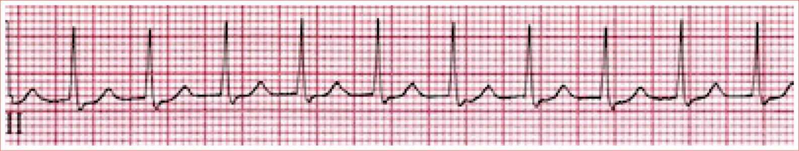
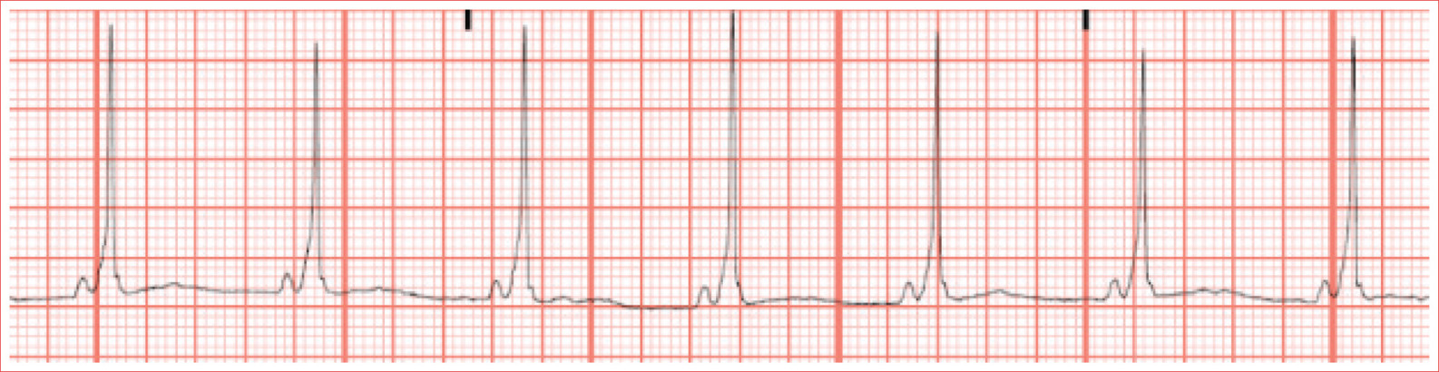
Normal Sinus Rhythm
Regular rhythm
Rate of 60 to 100 beats/minute
QRS complexes are typically less than 100 ms duration
P waves are present and ALWAYS upright in leads I and II and inverted in aVR
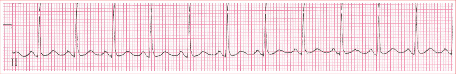
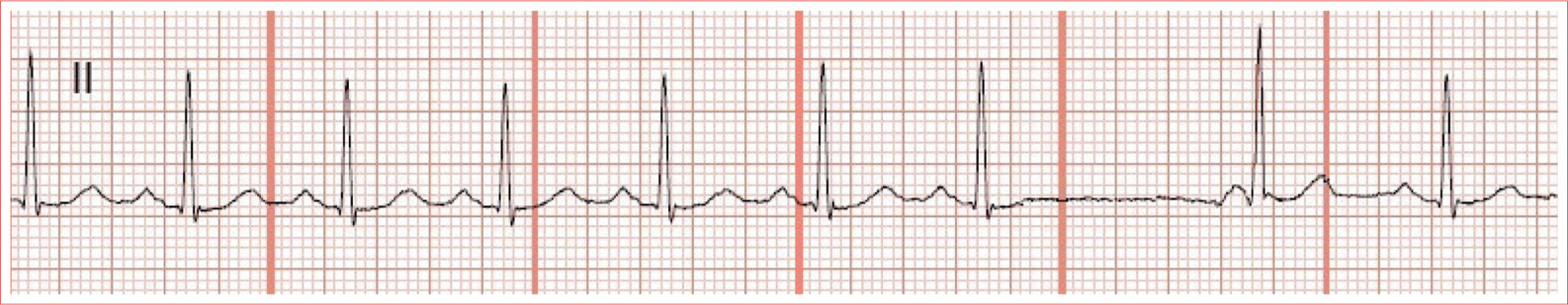
Sinus Bradycardia
Regular rhythm
Rate less than 60 beats/minute
QRS complexes are typically less than 100 ms duration
P waves upright in leads I and II and inverted in aVR
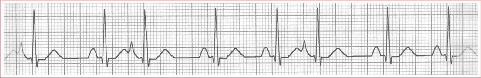
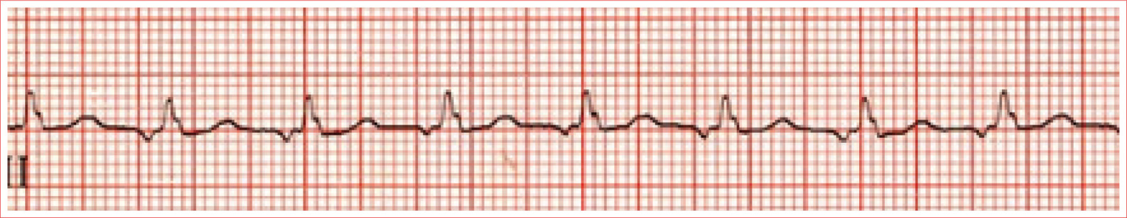
Sinus Tachycardia
Regular rhythm
Rate greater than 100 beats/minute
QRS complexes are typically less than 100 ms duration
P waves are upright in leads I and II and inverted in aVR
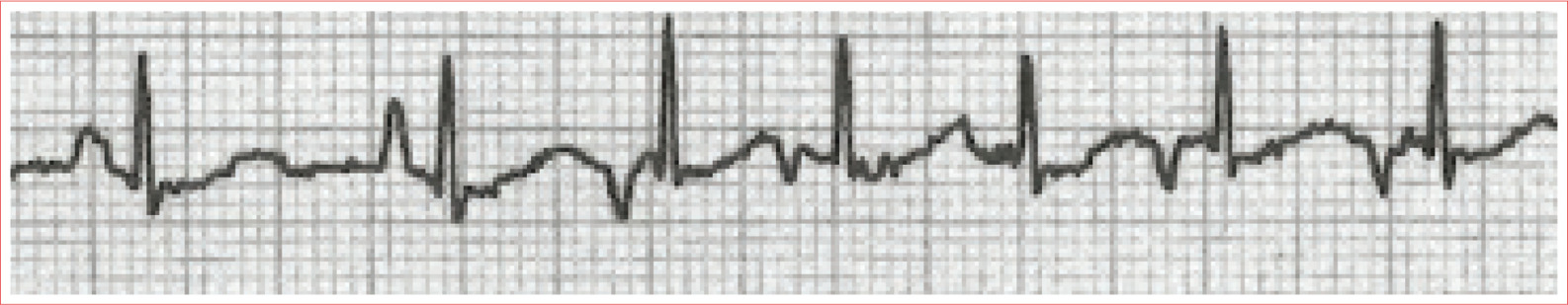
Sinus Arrhythmia
Irregular rhythm, P-P varies by more than 0.16 second
QRS complexes are typically less than 100 ms duration
P waves are upright in leads I and II and inverted in aVR
SUPRAVENTRICULAR ARRHYTHMIAS
Premature Atrial Complex
The P wave occurs before the next P wave was due
The P wave has a different appearance than the P wave
After the PAC, there is often a small pause before the SA node resumes control
The QRS complex is usually identical to all other QRS complexes
AV Nodal Reentry Tachycardia (AVNRT)
Regular rhythm
Rate usually 140 to 220 beats/minute
QRS is typically narrow
P waves are often hidden in the QRS complex
WolffParkinsonWhite Syndrome
Short PR interval
Delta wave; slurred upstroke to start QRS
Prolonged QRS complex greater than 0.11 second
ST-T-wave discordance
Pseudo-infarction pattern
Paroxysmal Ectopic Atrial Tachycardia
Regular rhythm
Rate greater than 100 beats/minute
QRS complex is typically narrow (<0.12 second)
P wave has a different morphology than the sinus beat
Isoelectric baseline
Often sudden in onset and sudden stop
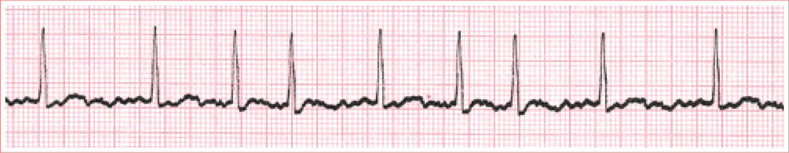
Multifocal Atrial Tachycardia/Wandering Atrial Pacemaker
Rhythm is irregularly irregular
MAT rate greater than 100 beats/minute
WAP rate less than 100 beats/minute
Narrow QRS complex
Three or more distinct P waves in a single lead
Isoelectric baseline
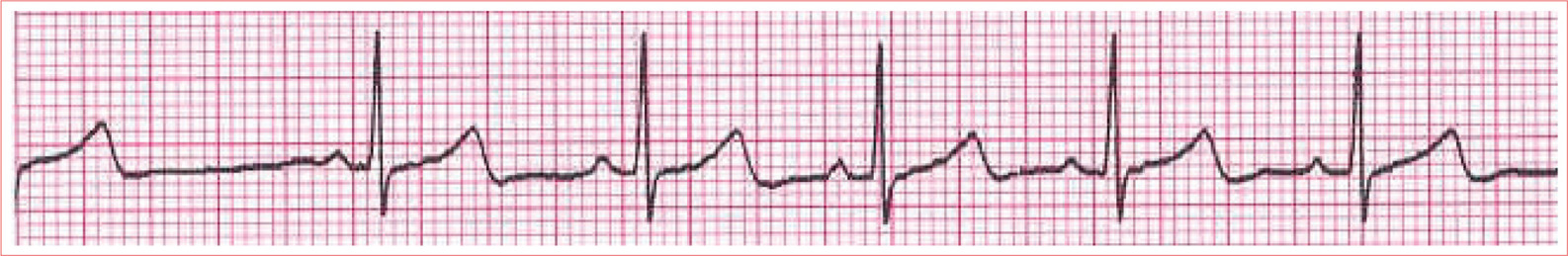
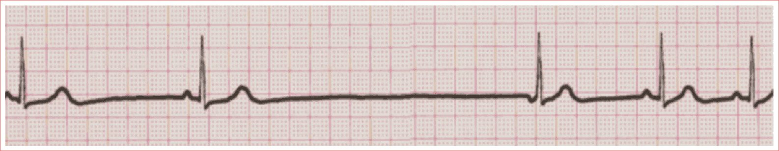
Atrial Escape Beat/Rhythm
Occurs after delay in sinus activity
P wave with different morphology than sinus beat, may be inverted
Rhythm is regular
Rate typically less than 60 beats/minute
Narrow QRS
PR interval greater than 0.12 second
Junctional Escape Beat/Rhythm
Occurs after delay in sinus activity
Rhythm is regular
Narrow QRS complex
P waves are absent or inverted immediately before or after QRS complex in lead II
PR is less than 0.12 second, when present
Atrial Fibrillation
Irregularly irregular ventricular response
Ventricular rate is commonly between 90 and 170 beats/minute
QRS complex is narrow
Absence of isoelectric baseline
No identifiable P waves
Atrial Flutter
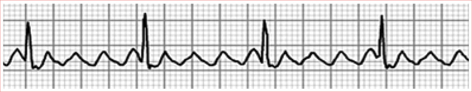
May have regular or irregular ventricular response
Ventricular rate is commonly 140 to 160 beats/minute, but variable
Ventricular rate is a ratio of atrial rate, most commonly a 2:1 AV ratio
Narrow QRS complex
Flutter or sawtooth waves
Absence of isoelectric baseline
VENTRICULAR ARRHYTHMIAS
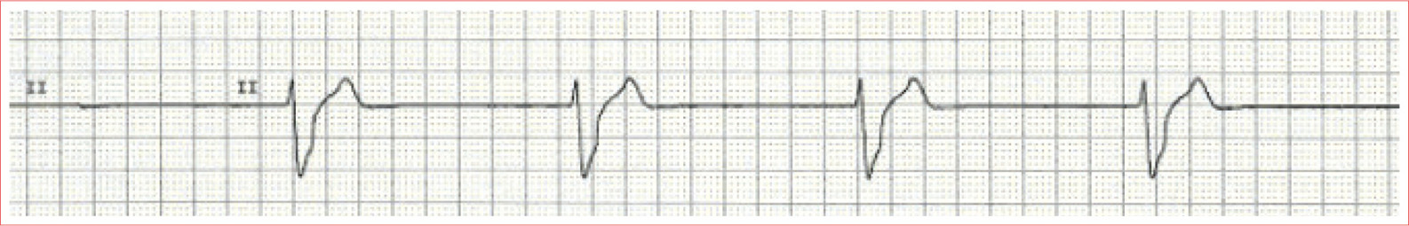
Premature Ventricular Complexes
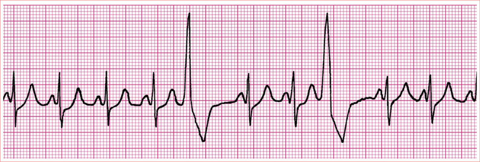
No apparent P wave
Premature QRS complex
Wide QRS (>0.12 second)
Bizarre
ST-T-wave discordance
Compensatory pause
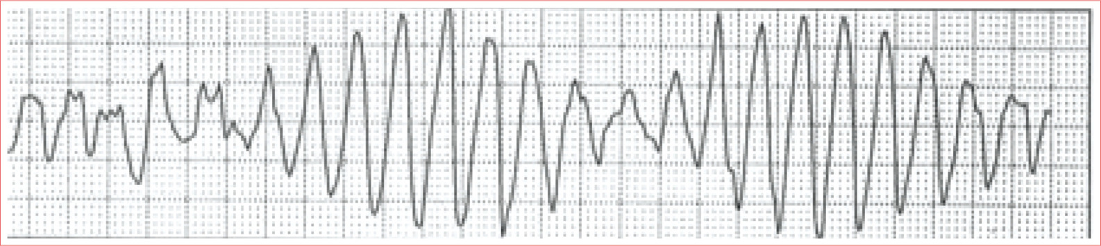
Monomorphic Ventricular Tachycardia
Three or more consecutive PVCs with same morphology
Regular rhythm
Rate greater than 120 beats/minute
Wide QRS
AV dissociation
Ventricular Fibrillation
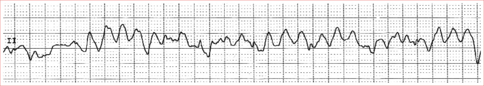
Fibrillatory waves
Irregular
Rate 150 to 500 quivers/minute
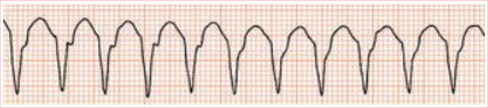
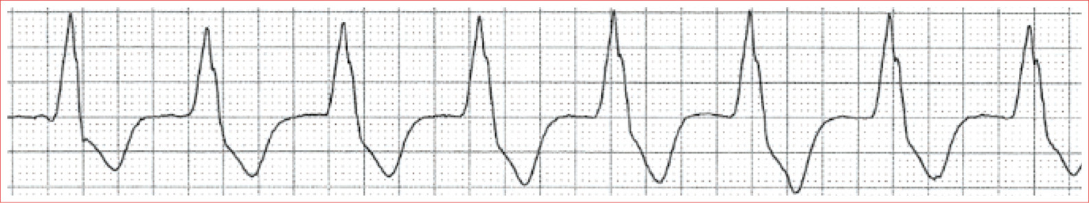
Torsades de Pointes
Irregular rhythm
Rate 150 to 200 beats/minute
Gradual shift in both axis and amplitude of QRS complexes
QT prolongation was cause
Ventricular Escape Rhythm
Regular rhythm
No P waves
Wide QRS
Rate is 20 to 50 beats/minute
Accelerated Idioventricular Rhythm
Regular rhythm
Absence of P waves
Next pageFont size:
Interval:
Bookmark:
Similar books «Mastering the 12-Lead EKG, Second Edition»
Look at similar books to Mastering the 12-Lead EKG, Second Edition. We have selected literature similar in name and meaning in the hope of providing readers with more options to find new, interesting, not yet read works.
Discussion, reviews of the book Mastering the 12-Lead EKG, Second Edition and just readers' own opinions. Leave your comments, write what you think about the work, its meaning or the main characters. Specify what exactly you liked and what you didn't like, and why you think so.