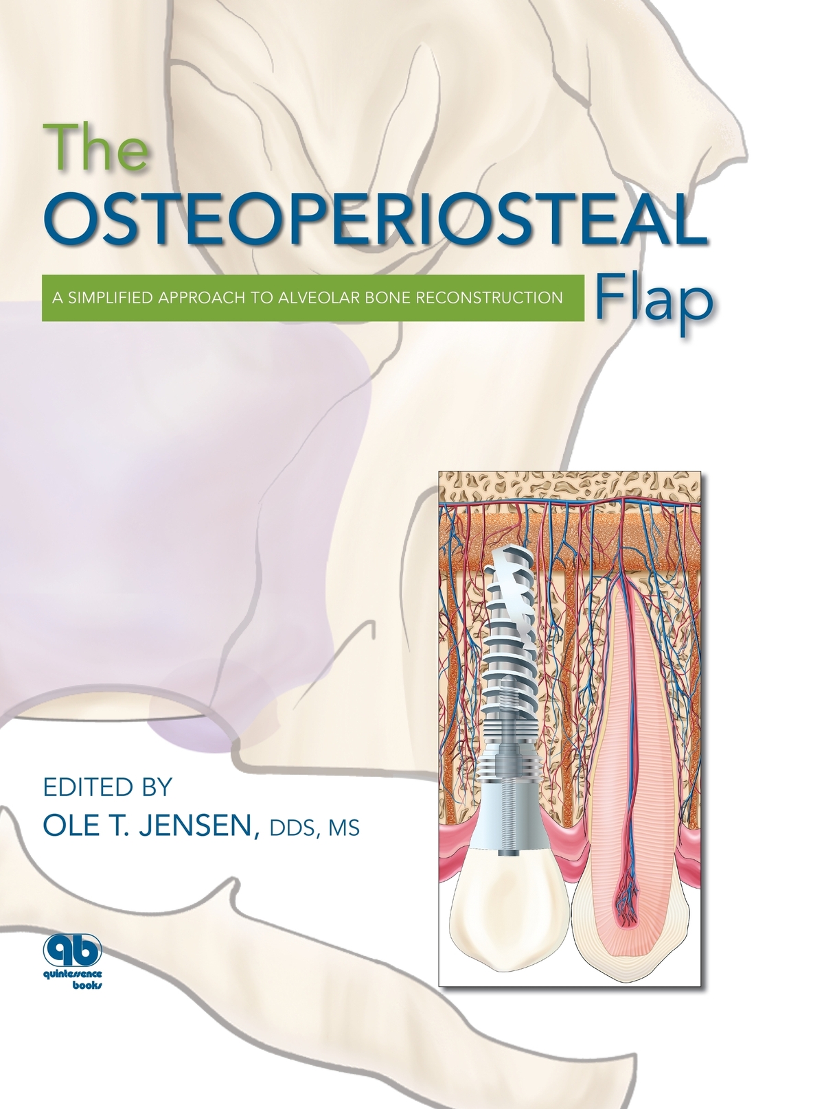
Ole T. Jensen - The Osteoperiosteal Flap: A Simplified Approach to Alveolar Bone Reconstruction
Here you can read online Ole T. Jensen - The Osteoperiosteal Flap: A Simplified Approach to Alveolar Bone Reconstruction full text of the book (entire story) in english for free. Download pdf and epub, get meaning, cover and reviews about this ebook. year: 2009, publisher: Quintessence Pub Co, genre: Home and family. Description of the work, (preface) as well as reviews are available. Best literature library LitArk.com created for fans of good reading and offers a wide selection of genres:
Romance novel
Science fiction
Adventure
Detective
Science
History
Home and family
Prose
Art
Politics
Computer
Non-fiction
Religion
Business
Children
Humor
Choose a favorite category and find really read worthwhile books. Enjoy immersion in the world of imagination, feel the emotions of the characters or learn something new for yourself, make an fascinating discovery.
- Book:The Osteoperiosteal Flap: A Simplified Approach to Alveolar Bone Reconstruction
- Author:
- Publisher:Quintessence Pub Co
- Genre:
- Year:2009
- Rating:3 / 5
- Favourites:Add to favourites
- Your mark:
The Osteoperiosteal Flap: A Simplified Approach to Alveolar Bone Reconstruction: summary, description and annotation
We offer to read an annotation, description, summary or preface (depends on what the author of the book "The Osteoperiosteal Flap: A Simplified Approach to Alveolar Bone Reconstruction" wrote himself). If you haven't found the necessary information about the book — write in the comments, we will try to find it.
Contents
Section I: Biologic Rationale
1. Biologic Basis of the Osteoperiosteal Flap
2. A New Biologic Classification of Bone Augmentation
Section II: Distraction Osteogenesis Techniques
3. Alveolar Distraction Osteogenesis
4. Supraperiosteal Transport Distraction Osteogenesis
5. Rapid Alveolar Expansion of Osteoperiosteal Flaps
Section III: Pedicled Segmental Osteotomy Techniques
6. Book Bone Flap
7. Island Osteoperiosteal Flap
8. Internal Alveolar Split Bone Graft
9. Sandwich Osteotomy Bone Graft in the Anterior Maxilla
10. Sandwich Osteotomy Combined with Extraction Socket Bone Graft
11. Sandwich Osteotomy Bone Graft in the Anterior Mandible
12. Smile Osteotomy
13. Sinus Graft Combined with Osteoperiosteal Flaps
14. Maxillary Alveolar Split Horseshoe Osteotomy
15. Sinus Floor Intrusion as a Vascularized Osteoperiosteal Flap
Section IV: Restorative Techniques
16. Alveolar Design by Stereolithography
17. Esthetically Driven Prosthetic Management of Osteoperiosteal Flaps
18. Esthetically Driven Surgical and Prosthetic Management of Alveolar Distraction Osteogenesis
19. Recombinant Protein Application for Bony and Periodontal Augmentation
20. Dental Implant Repositioning by Osteotomy in the Esthetic Zone
Section V: Developing Technologies
21. Osteoperiosteal Tissue-Engineered Injectable Bone
22. De Novo Tooth Engineering to Replace Lost Teeth
Ole T. Jensen: author's other books
Who wrote The Osteoperiosteal Flap: A Simplified Approach to Alveolar Bone Reconstruction? Find out the surname, the name of the author of the book and a list of all author's works by series.


The Osteoperiosteal Flap: A Simplified Approach to Alveolar Bone Reconstruction — read online for free the complete book (whole text) full work
Below is the text of the book, divided by pages. System saving the place of the last page read, allows you to conveniently read the book "The Osteoperiosteal Flap: A Simplified Approach to Alveolar Bone Reconstruction" online for free, without having to search again every time where you left off. Put a bookmark, and you can go to the page where you finished reading at any time.
Font size:
Interval:
Bookmark:

I would like to acknowledge my wife Marty, my children Sverre, Autumn, and Trygve, and my grandchildren as the underlying inspiration and drive for any success I might have outside the home.
I especially want to thank those who contributed so greatly to making this book possible: my publisher Tomoko Tsuchiya for taking a chance on me once again , Lisa Bywaters for her extreme patience and expert guidance, Bryn Goates for her positive and concise editing, Sue Robinson on an artistic layout and design, and Peter Jurek for his outstanding renderings.
I also want to express special thanks to Karen Shoop, my implant coordinator and brain away from home, Kristen Stifflear, who gave birth to a child this year and still provided many of the photos and essential content organization for the book, and my fantastic surgical assistants Cindy Formanek and Jennifer Patrick.
Dr Jared Cottam contributed as a research assistant; special thanks to him. Without my staff and associates, this decade-long process could not have been completed.
William H. Bell, DDS
To see what is in front of ones nose requires constant struggle.
George Orwell
In the early evolutionary years of oral and maxillofacial surgery, pulpal response to alveolar osteotomies was a central question to be answered. Relatively few surgeons, however, were interested in this fundamental question. At the annual association meetings of the American Association of Oral and Maxillofacial Surgeons or the International Association for Dental Research, it was not unusual to see only five or six surgeons in attendance in the research sessions debating the question of what constitutes a viable tooth.
It had been recognized for some time that teeth contained within a repositioned dento-osseous segment did not respond positively to electrical stimulation immediately after surgery. This aberrant testing was usually transient and results returned to normal after 3 to 8 months. A small, dedicated cadre of investigators16 often debated as to whether pulpal vascularity were more important than neuronal continuity.
In time, preservation of pulpal circulation was generally considered to be necessary if normal pulpal anatomy were to be preserved following dentoalveolar surgery. Neuronal, blood flow, and histologic studies gradually confirmed these findings and created enormous interest in the surgical repositioning of all maxillary and mandibular teeth by dentoalveolar surgery and orthodontics. These studies opened the gate to the possibility of simultaneous repositioning of all or a part of the maxilla and maxillary teeth independently as small dento-osseous segments.
Recent studies have used laser Doppler flowmetry to assess tooth vitality after Le Fort I osteotomy.7,8 These studies have clearly demonstrated that teeth without normal innervation can have an intact blood supply and be vital.
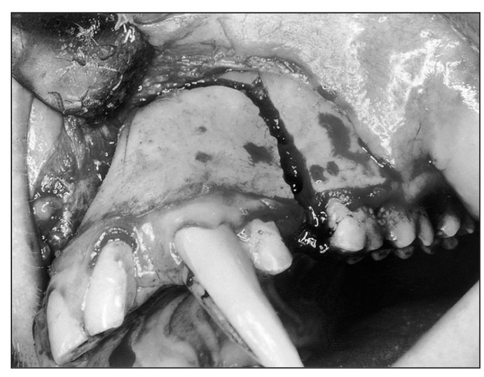
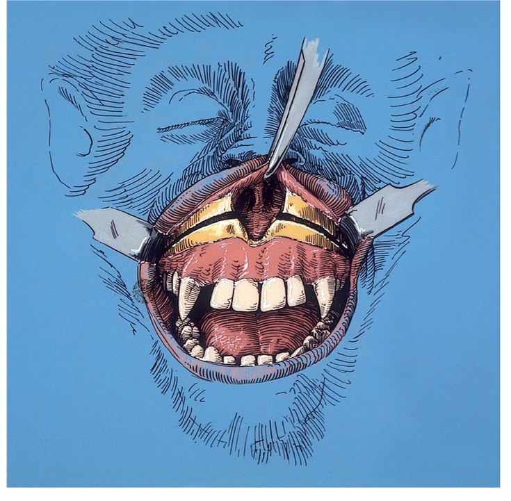
Fig 1-1a Anterior maxillary osteotomy performed after reflection of the labial and buccal mucoperiosteum. (Reprinted from Bell et al23 with permission.)
 Biology of Wound Healing
Maxillary deformities have been recognized and described for centuries, but the challenge to correct them through surgery in the anterior maxilla was not met until the turn of the century. Bold attempts to move the anterior maxilla were first made by Cohn-Stock,9 Wassmund,10 and Spanier,11 who were unaware of the biologic basis for the healing of such surgically created wounds. Analysis of Cohn-Stocks initial attempt to retroposition the anterior maxilla surgically indicates that he feared the consequences of such a procedure and attempted to avoid them by creating a greenstick fracture of the anterior maxilla through a transverse palatal incision; the retropo-sitioned maxilla subsequently relapsed.
When maxillary surgical procedures were introduced to the United States,1215 the rationale for use of the various surgical techniques for correcting dentofacial deformities was empirical.16 Basic questions concerning the healing of surgical wounds produced by maxillary osteotomies had not been investigated. Many surgeons believed that the maxilla healed by fibrous union. Others believed absolute stability was necessary. Devitalization of teeth and bone in the mobilized segments had been re-ported. Varying degrees of relapse subsequent to posterior maxillary osteotomy9 and total maxillary osteotomy were reported. The possibility that the maxilla could be successfully repositioned superiorly or inferiorly through surgery was doubted by many clinicians and scientists. The blood vessels necessary to maintain circulation to the mobilized bony segments and teeth had not been studied. Consequently, both one-stage and two-stage procedures (of empirical duration ranging between 2 and 8 weeks) were devised to prevent impairment of the vascular supply to the mobilized dentoalveolar segments.17
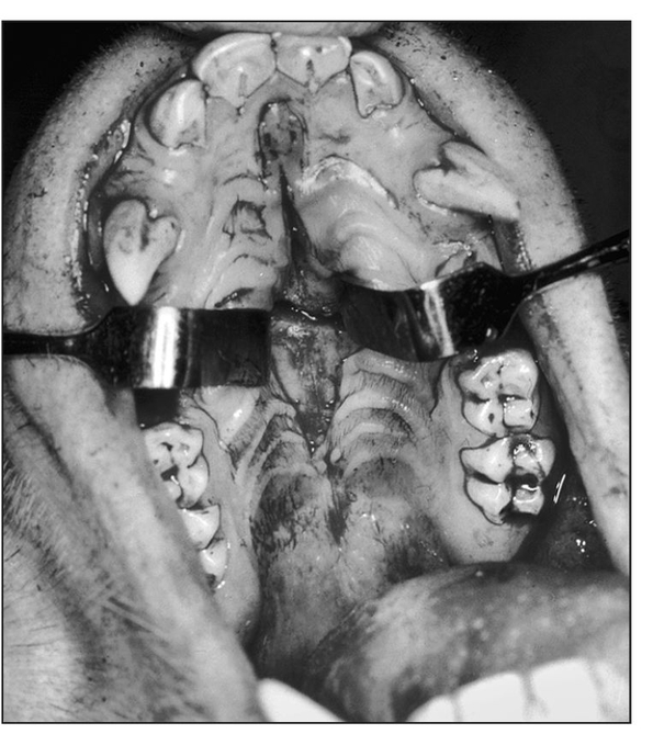
Fig 1-1b Midpalatal sagittal incision for palatal osteotomies. (Reprinted from Bell et al23 with permission.)
In 1962, animal and clinical investigations were initiated to delineate the biology of maxillary osteotomy wound healing. Since then, rabbits, dogs, monkeys, and baboons have been used as experimental models to investigate the revascularization and bone healing associated with various maxillary techniques.12,16,18,19 Macaca mulatta was usually selected as the experimental animal of choice because of its anatomic, physiologic, and dental similarities to the human. Because maxillary osteotomies are usually performed in adults, large male rhesus monkeys from 8 to 14 years of age and weighing an average of 9 kg, were chosen for study.
From 1962 to 1965, revascularization and bone healing were studied on animal models after clinical simulations of three variations of anterior maxillary osteotomy techniques10,14,20 (Fig 1-1) were performed via various flap designs to validate vascularity to the repositioned osseous segments.12,13,22 The animals were killed 1, 3, 6, and 24 weeks after surgery for microangiographic and histologic investigation.
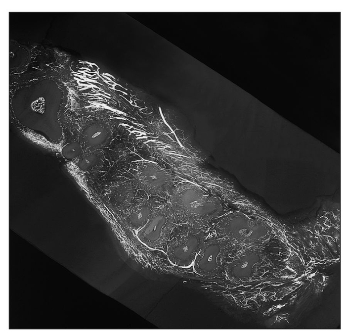
Horizontal microangiogram demonstrating the vascular pattern of a control animal. A reticulated network of periodontal plexus encircles each tooth, composed of anastomosing blood vessels from the labial (facial), gingival, intra-alveolar, and apical vessels. (Reprinted from Bell et al21 with permission.)
Serial 1-mm transverse, sagittal, and horizontal tissue slices were cut from the specimens for microangiographic study, which were in turn cut into seven microscopic slices for histologic study. Microangiographic and histologic techniques demonstrated that intraosseous and intrapulpal circulation to the anterior maxillary segment was maintained when soft tissue was kept intact.12,22 Osteonecrosis was minimal and vascular ischemia was only transient when the anterior maxillary bone segment was pedicled to the labiobuccal mucoperiosteum, palatal mucoperiosteum (), or a combination of both. Osseous union between most of the sectioned segments occurred within 6 weeks without immobilization of the mandible.
Font size:
Interval:
Bookmark:
Similar books «The Osteoperiosteal Flap: A Simplified Approach to Alveolar Bone Reconstruction»
Look at similar books to The Osteoperiosteal Flap: A Simplified Approach to Alveolar Bone Reconstruction. We have selected literature similar in name and meaning in the hope of providing readers with more options to find new, interesting, not yet read works.
Discussion, reviews of the book The Osteoperiosteal Flap: A Simplified Approach to Alveolar Bone Reconstruction and just readers' own opinions. Leave your comments, write what you think about the work, its meaning or the main characters. Specify what exactly you liked and what you didn't like, and why you think so.