Papertowns Publishers
72, Vishwanath Dham Colony,
Niwaru Road, Jhotwara,
Jaipur, 302012
A Simple Approach to Neuroscience
Copyright Abhijeet Satani, 2020
ISBN Print Book - 978-93-87131-59-0
All rights reserved. No part of this book may be reproduced or transmitted in any form or by any means, electronic or mechanical, including photocopying, recording, or by an information storage and retrieval systemexcept by a reviewer who may quote brief passages in a review to be printed in a magazine, newspaper, or on the Webwithout permission in writing from the copyright owner.
Although the author and publisher have made every effort to ensure the accuracy and completeness of information contained in this book, we assume no responsibility for errors, inaccuracies, omissions, or any inconsistencies herein. Any slights on people, places, or organizations are unintentional.
Printed in India
No diet will remove all the fat from your body, because the brain is entirely fat. Without a brain, you might look good, but all you could do is run for public office.
George Bernard Shaw
The human brain is the most complex organ, and arguably the most complex thing on earth. This booklet is an introduction for young students about neuroscience.
The course has been designed for beginners in Neuroscience. The course has been designed in a way that it suits the requirements of all, concerned with the study of Neuroscience and Neurology, Brain and its correlates.
The concepts and terminology of neuroscience have been presented in simple, and easy to understand language to facilitate the beginners of Brain and Brain Sciences.
New techniques such as electrodes that can touch the surface of cells, human brain scanning machines and touch on some of the ethical issues and social implications emerging from brain research.
Chapter 1
Anatomy of the Brain
The brain gives meaning to the things around us and handles essential functions in the human body. We have 5 sense, namely smell, sight, touch, taste and hearing, through which the brain gathers many messages at a time. It controls functions of most organs of the body, such as the movement of the limbs, thoughts, memory, speech, etc. It also handles reactions to stressful situations like anxiety, illness, childbirth, etc. by regulations heart and breathing rates. The many components of the brain perform very specific and important functions.
The weight of the brain is not constant. At birth, it weighs about one pound on average and grows a pound bigger during childhood. At adulthood, the average female brain weighs about 2.7 pounds, while an average male brain weighs about 3 pounds.
The nervous system
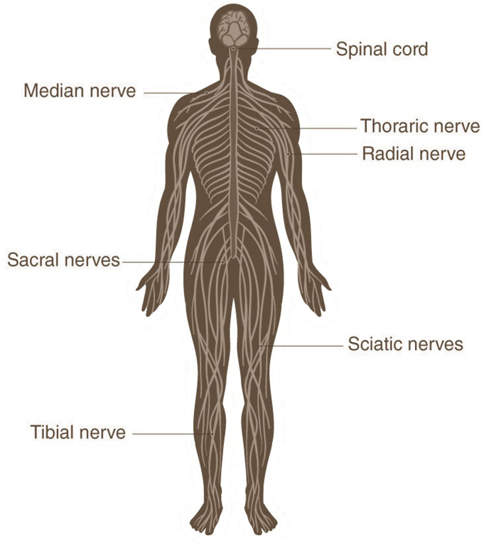
The nervous system is divided into two parts, namely the central nervous system and the peripheral nervous system. The central nervous system consists of the brain, the cranial nerves and the spinal cord. The peripheral nervous system is made up of the spinal nerves that stem from the spinal cord and the autonomous nervous system that is further divided into the sympathetic and the parasympathetic nervous system.
The cell structure of the brain
The brain is composed of two types of cells, namely neurons and glial cells, or as is also known as neuroglia or glia. The job of a neuron is to send and receive impulses or signals. Glial cells, on the other hand, supply support and nutrition, maintain homeostasis, form myelin and facilitate the process of signal transmission in the nervous system. The ratio of glial cells to neurons is 50:1.
In primary brain tumors, glial cells are the most common. When a person has a brain tumor, a biopsy is done and brain tissue is removed from the tumor for identification by a pathologist. The pathologist identifies the type of cells present in this tissue and tumors are thus named based on this connection. The type of brain tumor and cells comprised in this impact patients prognosis and treatment.
The Meninges
The brain is safeguarded by the bone structure called the cranium. It protects the brain from physical trauma and injuries. Along with the cranium, the bones that form and protect the face are together called the skull. Between the skull and the brain is 3 layers of tissue that covers the brain and the spinal cord called the Meninges. The three layers are called the Dura mater, Arachnoid and Pia mater from the outer layer to the middle layer to innermost layer respectively.
Dura Mater
The Dura mater is further made of two layers of whitish, inelastic membrane. The outer membrane is called the Periosteum, while the inner membrane is called the Dura. This Dura runs through the entire skull and folds in places to create pockets in which parts of the brain are protected. The falx is one such fold. It separates the right and left half of the brain. Another fold is the tentorium that separates the upper and the lower brain parts.
Arachnoid
The Arachnoid is the central layer of the meninges. This is thin and fragile and it covers the entire brain. The Arachnoid is formed of delicate elastic tissues and blood vessels of different sizes. The space between the Dura and the Arachnoid membranes is called the subdural space.
Pia Mater
This is the last layer of meninges and it is closed to the brain surface. This membrane has many blood vessels that penetrate deep into the brain surface. The Pia mater covers the entire brain while also following the folds of the brain. The major arteries provide Pia with its blood vessels to supply blood and nutrients to the brain for functioning. The space separating the Arachnoid and the Pia is called the subarachnoid space. Within this area, cerebrospinal fluid flows.
Cerebrospinal Fluid
Cerebrospinal fluid, or CSF for short, is present within the brain and it covers the brain and the spinal cord. It acts as a cushion for the brain and the spine, protecting them from injury. It is clear in color and watery. This fluid is continuously being absorbed and replenished while it is circulating around the brain and the spinal cord in channels. Within hollow conduits in the brain, this fluid is produced. These conduits are called the ventricles, and a structure within each ventricle specifically responsible for the production of this CSF is called the Choroid Plexus. Even though there is a balance between the production and absorption of CSF, disruptions are possible.
The Ventricular System
A series of foramen (holes) and tubes connect the four ventricles of the ventricular system. The ventricles are like cavities, two of which are called the lateral ventricles and they are walled in the cerebral hemispheres. They both communicate with the third ventricle via openings called Foramen of Munro. The openings are separate for both the lateral ventricles. This third ventricle is located in the center of the brain with walls composed of thalamus and hypothalamus, and it connects with the fourth ventricle via a long tube called the Aqueduct of Sylvius. The CSF flows through the fourth ventricle and passes through a series of openings to flow around the brain and the spinal cord.


