Pearce Wilcox , Jeremy Road and Ali Altalag Pulmonary Function Tests in Clinical Practice 10.1007/978-1-84882-231-3_1 Springer-Verlag London Limited 2009
1. Spirometry
Ali Altalag 1, Jeremy Road 2 and Pearce Wilcox 3
(1)
University of Toronto, 23 lvor Rd.,, M4N 2H3 North York, Ontario, Canada
(2)
The Lung Centre, Vancouver General Hospital, 7th Floor, 2775 Laurel Streen, V5Z 1M9 Vancouver, BC, Canada
(3)
Cpy Pauls Hospital, 1081 Burrard Street, V6Z 1Y6 Vancouver, BC, Canada
Abstract
Spirometry is the most essential part of any pulmonary function study and provides the most information. In spirometry, a machine called a spirometer is used to measure certain lung volumes, called dynamic lung volumes. The two most important dynamic lung volumes measured are the forced vital capacity (FVC) and the forced expiratory volume in the 1st second (FEV1). This section deals with the definitions of these and other terms.
Spirometry is the most essential part of any pulmonary function study and provides the most information. In spirometry, a machine called a spirometer is used to measure certain lung volumes, called dynamic lung volumes. The two most important dynamic lung volumes measured are the forced vital capacity (FVC) and the forced expiratory volume in the 1st second (FEV1). This section deals with the definitions of these and other terms.
DEFINITIONS
Forced Vital Capacity
Is the volume of air in liters that can be forcefully and maximally exhaled after a maximal inspiration. FVC is unique and reproducible for a given subject.
The slow vital capacity (SVC) also called the vital capacity (VC) is similar to the FVC, but the exhalation is slow rather than being as rapid as possible as in the FVC. In a normal subject, the SVC usually equals the FVC,.
Figure 1.1
FVC and SVC are compared with each other in a normal subject ( a ) and in a patient with an obstructive disorder ( b ). In case of airway obstruction, SVC is larger than FVC, indicating air trapping
Table 1.1
Definitions of obstructive and restrictive disorders
Obstructive disorders |
Are characterized by diffuse airway narrowing secondary to different mechanisms [immune related, e.g., bronchial asthma, or environmental, e.g., chronic obstructive pulmonary disease (COPD)] |
Restrictive disorders |
Are a group of disorders characterized by abnormal reduction of the lung volumes, either because of alteration in the lung parenchyma or because of a disease of the pleura, chest wall or due to muscle weakness |
The inspiratory vital capacity (IVC) is the VC measured during inspiration rather than expiration. The IVC should equal the expiratory VC. If it does not, poor effort or an air leak could be responsible. IVC may be larger than the expiratory VC in patients with significant airway obstruction, as in this case the increased negative intrathoracic pressure opens the airways facilitating inspiration, as opposed to the narrowing of airways during exhalation as the intrathoracic pressure becomes positive. Narrowed airways reduce airflow and hence the amount of exhaled air.
Forced Expiratory Volume in the 1st Second
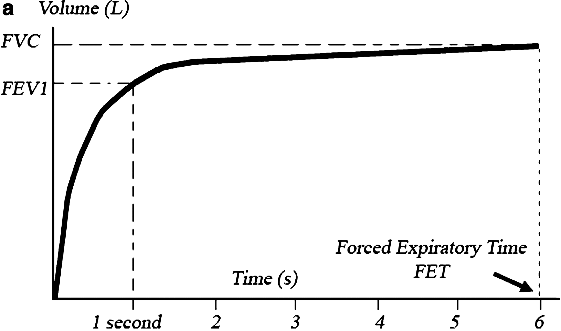
Is the volume of air in liters that can be forcefully and maximally exhaled in the 1st second after a maximal inspiration. In other words, it is the volume of air that is exhaled in the 1st second of the FVC, and it normally represents 80% of the FVC.
FEV is similarly defined as the volume of air exhaled in the first 6 s of the FVC and its only significance is that it can sometimes substitute the FVC in patients who fail to exhale completely.
FEV1/FVC Ratio
This ratio is used to differentiate obstructive from restrictive lung disorders; see Table for definitions. In obstructive disorders, FEV1 drops much more significantly than FVC and the ratio will be low, while in restrictive disorders, the ratio is either normal or even increased as the drop in FVC is either proportional to or more marked than the drop in FEV1.
Normally, the FEV1/FVC ratio is greater than 0.7, but it decreases (to values <0.7) with normal aging. The changes in the elderly probably reflect the decrease in elastic recoil of the lungs that occurs with aging.
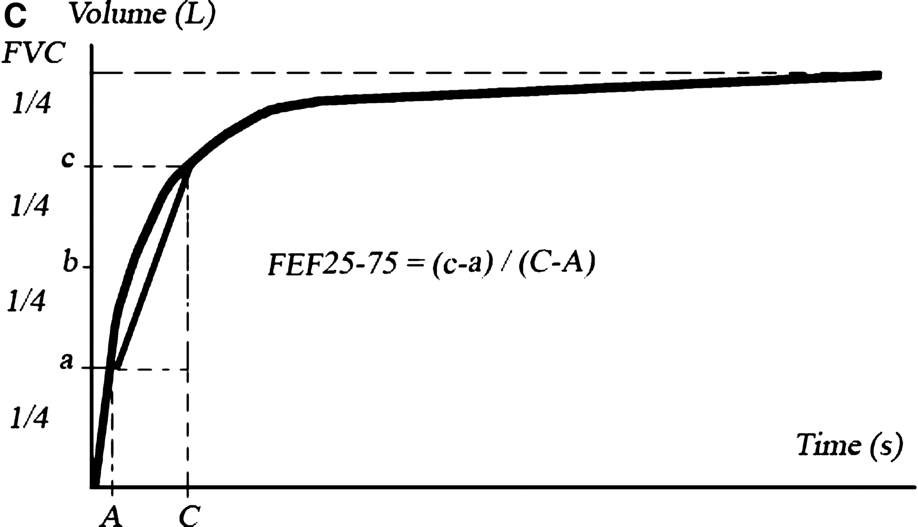
The Instantaneous Forced Expiratory Flow (FEF25, FEF50, FEF75) and the Maximum Mid-Expiratory Flow (MMEF or FEF2575)
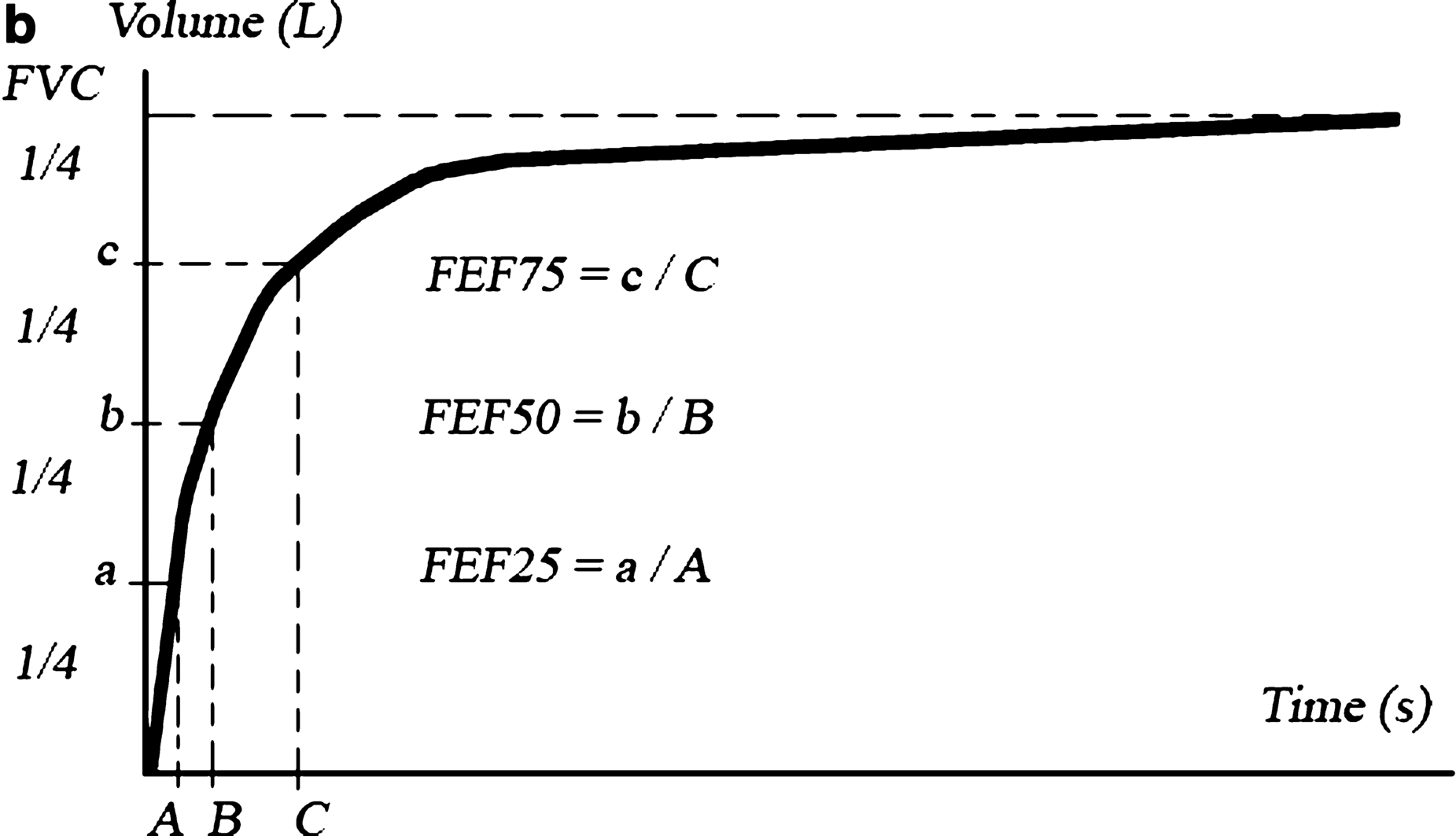
The instantaneous forced expiratory flow (FEF) represents the flow of the exhaled air measured (in liters per second) at different points of the FVC, namely at 25, 50, and 75% of the FVC. They are abbreviated as FEF , FEF , and FEF , respectively; Fig.
Figure 1.2
The volumetime curve (spirogram). The following data can be acquired: ( a ) FVC is the highest point in the curve; FEV1 is plotted in the volume axis opposite to the point in the curve corresponding to 1 s; duration of the study (the forced expiratory time or FET) can be determined from the time axis, 6 s in this curve. ( b ) FEF25,50,75 can be roughly determined by dividing the volume axis into four quarters and determining the corresponding time for each quarter from the time axis. Dividing the volumes (a, b, and c) by the corresponding time (A, B, and C) gives the value of each FEF (FEF25, FEF50, FEF75, respectively). Note that this method represents a rough determination of FEFs, as FEFs are actually measured instantaneously by the spirometer and not calculated. ( c ) FEF2575 can be roughly determined by dividing the volume during the middle half of the FVC (ca) by the corresponding time (CA). FEF2575 represents the slope of the curve at those two points
Peak Expiratory Flow
Is the maximum flow (in liters per second) of air during a forceful exhalation. Normally, it takes place immediately after the start of the exhalation and it is effort-dependent. PEF drops with a poor initial effort and in obstructive and, to a lesser extent, restrictive disorders. PEF measured in the laboratory is similar to the peak expiratory flow (PEF) rate (in liters per minute) that is measured routinely at the bedside to monitor asthmatic patients.
SPIROMETRIC CURVES
The VolumeTime Curve (The Spirogram)
Is simply the FVC plotted as volume in liters against time in seconds; Fig..
You can extract from this curve both the FVC and FEV1. FEV1/FVC ratio can be estimated by looking at where the FEV1 stands in relation to the FVC in the volume axis; Figure. , c.


