James A. Nunley (ed.) The Achilles Tendon Treatment and Rehabilitation 10.1007/978-0-387-79205-7_1 Springer Science+Business Media, LLC 2008
1. Anatomy of the Achilles Tendon
Florian Nickisch 1
(1)
University of Utah Orthopaedic Center, 590 Wakara Way, Salt Lake City, UT 84108, USA
The Achilles tendon is the conjoined tendon of the two heads of the gastrocnemius and the soleus muscle. Together these structures are often referred to as the gastroc-soleus complex. It is the largest and strongest tendon in the human body and subject to tensile forces of up to 12.5 times body weight (9 kilonewton [kN]) during sprinting Due to its size and functional demands, the Achilles tendon is susceptible to both acute and chronic injuries and is directly or indirectly implicated in many pathologic conditions of the foot and ankle. To diagnose and treat these disorders, a thorough knowledge of the anatomy of the Achilles tendon and its surrounding structures is crucial.
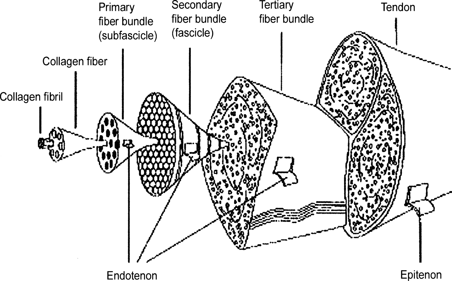
Microstructure
Tendons are complex, composite, roughly uniaxial structures consisting of collagen fibrils embedded in a matrix, rich in water and proteoglycans with a paucity of cells. Collagen (mainly type I collagen) accounts for 65% to 80%, and elastin for approximately 2%, of the dry mass of the tendon. The predominant cells are tenoblasts and tenocytes (elongated fibroblasts). Their spindle-shaped cell bodies are arranged in rows between the collagen fiber bundles and they produce the extracellular matrix proteins. Soluble tropocollagen molecules are cross-linked to create insoluble collagen molecules, which then aggregate into microfibrils. Further aggregation leads to the formation of collagen fibrils. The diameter of collagen fibrils in the Achilles tendon varies from 30 nm to 150 nm.
Figure 1.1.
The organization of tendon structure from collagen fibrils to the entire tendon. (From Kannus, with permission from Blackwell Publishing.)
Gross Anatomy
The leg consists of four compartments (anterior, lateral, superficial, and deep posterior) divided by strong fascial septa. The superficial posterior compartment contains the gastroc-soleus complex and the plantaris muscle, supplied by the tibial nerve and branches from the posterior tibial and peroneal arteries. It is separated from the deep posterior compartment by the deep leaf of the fascia cruris.
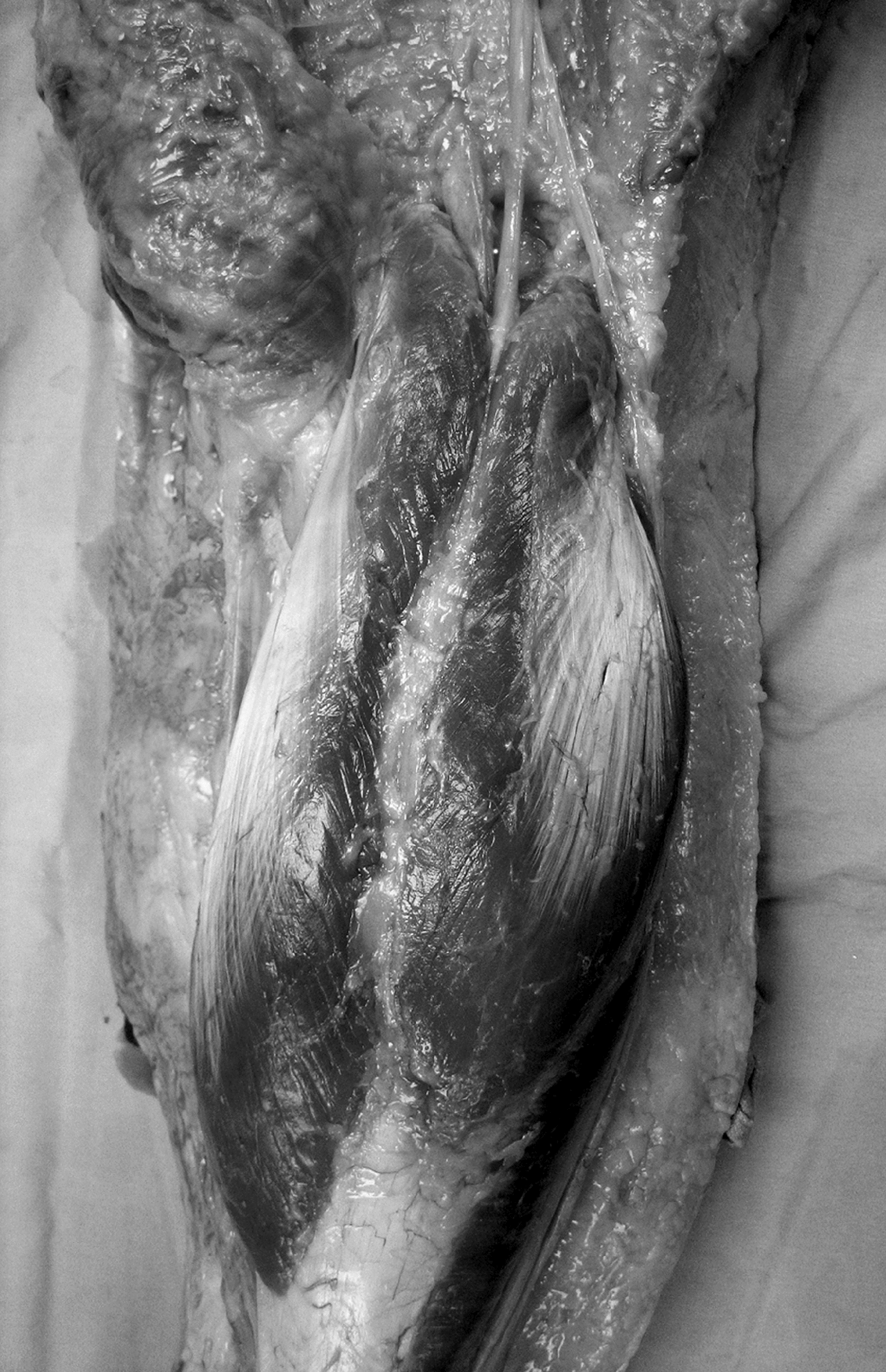
Gastrocnemius
The gastrocnemius muscle crosses the knee, ankle, and subtalar joint; hence, it is maximally stretched with the knee fully extended and the ankle dorsiflexed while the heel is inverted. When present, it is usually bilateral and serves as an attachment site for the fabellofibular ligament in the posterolateral corner of the knee joint capsule. The muscle fibers from each head run obliquely and attach at an angle in the middle of the calf into a midline raphe that further distal broadens into an aponeurosis on the anterior surface of the muscle. This aponeurosis gradually narrows and unites with the tendon of the soleus to form the Achilles tendon. The gastrocnemius is innervated by the first and second sacral roots through the tibial nerve.
Figure 1.2.
Posterior view of the gastrocnemius muscle and popliteal fossa. The common peroneal nerve passes just lateral to the lateral head, whereas the tibial neurovascular bundle passes between the medial and lateral heads of the muscle.
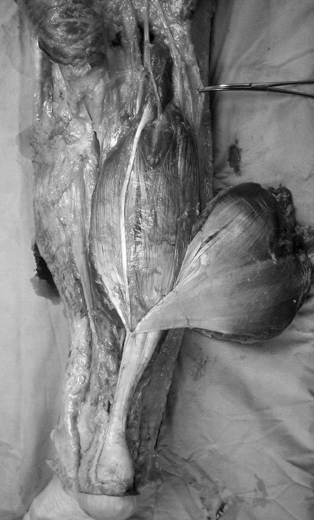
Soleus
The soleus is a postural muscle consisting mainly of slow-twitch muscle fibers. It helps to keep the body upright in stance and prevents the body from falling forward during gait, as it contracts when the center of gravity passes in front of the knee joint. It is the strongest muscle in the lower leg and the prime plantar flexor of the ankle joint. The gastrocnemius is innervated by the first and second sacral roots through the tibial nerve.
Figure 1.3.
Posterior view of the soleus muscle. The gastrocnemius has been detached at its origin and reflected laterally, exposing the plantaris muscle and tendon.
An accessory soleus muscle has been recognized since the 19th century. Originally thought to be a rare finding, it has been diagnosed more frequently since the introduction of magnetic resonance imaging (MRI) into clinical practice.
Plantaris
The plantaris has it origin on the lower part of the lateral prolongation of the linea aspera, and on the oblique popliteal ligament of the posterolateral knee joint capsule. It has a small fusiform, usually a 7- to 10-cm-long muscle belly. The thin plantaris tendon crosses obliquely between the gastrocnemius and soleus muscles and then runs parallel to the medial aspect of the Achilles tendon (Fig. Occasionally, the tendon is lost in the laciniate ligament or in the fascia of the leg. The plantaris is absent in 6% to 8% of individuals.
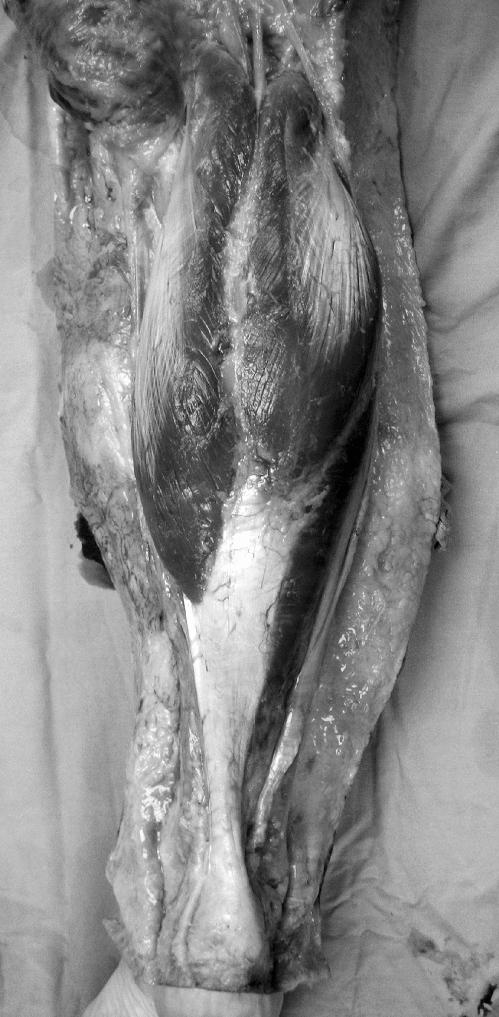
Achilles Tendon
The Achilles tendon originates in the middle of the lower leg as the confluence of the tendons of the gastrocnemius and soleus muscles at the gastroc-soleus junction (Figs.
Figure 1.4.
Posterior view of the Achilles tendon.
Muscle fibers of the soleus may insert into the anterior surface of the tendon to almost its insertion. The contribution of fibers of the gastrocnemius and soleus to the Achilles tendon is variable. In most individuals, the soleus contributes more fibers than the gastrocnemius, as demonstrated by Cummins and coworkers Fiber rotation reaches a maximum 2 to 5 cm proximal to the tendon insertion and creates high stresses in this area of the tendon, which may explain the poor vascularity and susceptibility to degeneration and injury in this region.
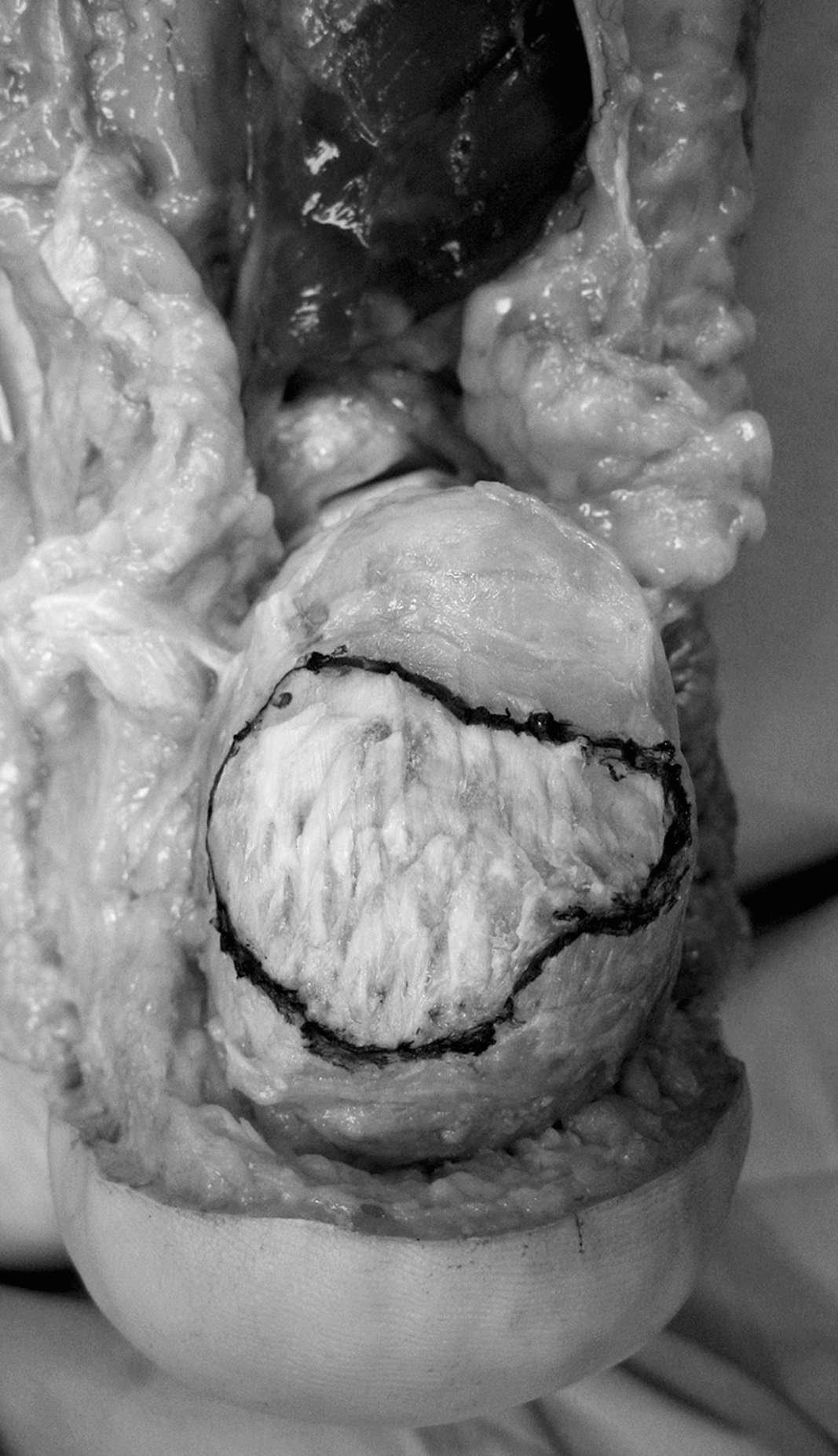
The Achilles tendon inserts on the middle third of the posterior surface of the calcaneal tuberosity, starting approximately 1 cm distal to the most superior border of the bone (Fig.
Figure 1.5.
Achilles tendon insertion on the posterior aspect of the calcaneal tuberosity. Note the broader area of insertion on the medial aspect as well as its central location on the calcaneal tuberosity.
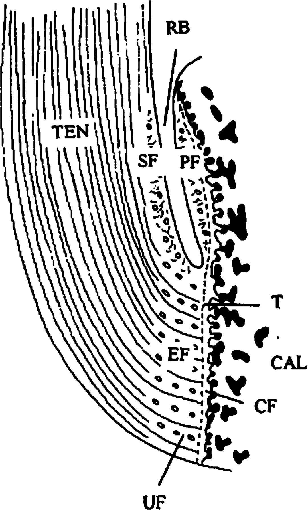
The attachment site of the Achilles tendon displays the typical structure of a fibrocartilaginous enthesis, and thus four zones of tissue are commonly present: pure dense fibrous connective tissue, uncalcified fibrocartilage, calcified fibrocartilage, and bone (Fig.
Figure 1.6.
A diagrammatic representation of the attachment of the human Achilles tendon (TEN) to the calcaneus (CAL). Note the three fibrocartilages at the insertion site: Enthesial fibrocartilage (EF) at the bonetendon junction; sesamoid fibrocartilage (SF) on the adjacent, deep surface of the tendon; and periosteal fibrocartilage (PF) on the opposing surface of the calcaneus. SF and PF are separated by the retrocalcaneal bursa (RF), but they are pressed against each other during ankle movements. EF has a zone of uncalcified fibrocartilage (UF), which is separated from a zone of calcified fibrocartilage (CF) by a tidemark (T), indicated by the broken line. (From Waggett et al., with permission from Elsevier.)