• Complain

Mehmet Turgut - The Sutures of the Skull: Anatomy, Embryology, Imaging, and Surgery

Here you can read online Mehmet Turgut - The Sutures of the Skull: Anatomy, Embryology, Imaging, and Surgery full text of the book (entire story) in english for free. Download pdf and epub, get meaning, cover and reviews about this ebook. year: 2021, publisher: Springer, genre: Romance novel. Description of the work, (preface) as well as reviews are available. Best literature library LitArk.com created for fans of good reading and offers a wide selection of genres:

Romance novel Science fiction Adventure Detective Science History Home and family Prose Art Politics Computer Non-fiction Religion Business Children Humor

Choose a favorite category and find really read worthwhile books. Enjoy immersion in the world of imagination, feel the emotions of the characters or learn something new for yourself, make an fascinating discovery.

Mehmet Turgut The Sutures of the Skull: Anatomy, Embryology, Imaging, and Surgery

The Sutures of the Skull: Anatomy, Embryology, Imaging, and Surgery: summary, description and annotation

We offer to read an annotation, description, summary or preface (depends on what the author of the book "The Sutures of the Skull: Anatomy, Embryology, Imaging, and Surgery" wrote himself). If you haven't found the necessary information about the book — write in the comments, we will try to find it.

This book provides an in-depth review of the sutures of the skull. The premature closure of the sutures of the skull (craniosynostosis) due to genetic or metabolic etiologies results in typical progressive skull deformity, due to both the inhibition of growth caused by the affected cranial suture and associated compensatory expansion of the skull along the open ones. Today, it is well known that early diagnosis of craniosynostosis is crucial for the best surgical outcomes and for the normal development of the brain and cosmetic appearance of the skull. As such, in addition to the anatomy, biology, genetics and embryology of the sutures of the skull, the book also covers the diagnosis and treatment of different forms of craniosynostosis such as metopism, and animal models for cranial suture research. This comprehensive work is a valuable resource for neuroscientists at all levels, from graduate students to researchers, as well as neurosurgeons, neuroanatomists, pediatricians, and neurologists seeking both basic and more advanced information on the unique structure of the sutures of the human skull.

Mehmet Turgut: author's other books


Who wrote The Sutures of the Skull: Anatomy, Embryology, Imaging, and Surgery? Find out the surname, the name of the author of the book and a list of all author's works by series.

The Sutures of the Skull: Anatomy, Embryology, Imaging, and Surgery — read online for free the complete book (whole text) full work

Below is the text of the book, divided by pages. System saving the place of the last page read, allows you to conveniently read the book "The Sutures of the Skull: Anatomy, Embryology, Imaging, and Surgery" online for free, without having to search again every time where you left off. Put a bookmark, and you can go to the page where you finished reading at any time.

Light

Font size:

Reset

Interval:

Bookmark:

Make
Contents
Landmarks
Book cover of The Sutures of the Skull Editors Mehmet Turgut R Shane - photo 1
Book cover of The Sutures of the Skull
Editors
Mehmet Turgut , R. Shane Tubbs , Ahmet T. Turgut and Aaron S. Dumont
The Sutures of the Skull
Anatomy, Embryology, Imaging, and Surgery
1st ed. 2021
Logo of the publisher Editors Mehmet Turgut School of Medicine - photo 2
Logo of the publisher
Editors
Mehmet Turgut
School of Medicine, Department of Neurosurgery, Aydn Adnan Menderes University, Efeler, Aydin, Turkey
R. Shane Tubbs
Departments of Neurosurgery, Neurology, Surgery, and Structural and Cellular Biology, Tulane University School of Medicine, Center for Clinical Neurosciences, New Orleans, LA, USA
Ahmet T. Turgut
Department of Radiology, Yksek htisas University, Ankara, Turkey
Aaron S. Dumont
Department of Neurosurgery, Tulane University, New Orleans, LA, USA
ISBN 978-3-030-72337-8 e-ISBN 978-3-030-72338-5
https://doi.org/10.1007/978-3-030-72338-5
The Editor(s) (if applicable) and The Author(s), under exclusive license to Springer Nature Switzerland AG 2021
This work is subject to copyright. All rights are solely and exclusively licensed by the Publisher, whether the whole or part of the material is concerned, specifically the rights of translation, reprinting, reuse of illustrations, recitation, broadcasting, reproduction on microfilms or in any other physical way, and transmission or information storage and retrieval, electronic adaptation, computer software, or by similar or dissimilar methodology now known or hereafter developed.
The use of general descriptive names, registered names, trademarks, service marks, etc. in this publication does not imply, even in the absence of a specific statement, that such names are exempt from the relevant protective laws and regulations and therefore free for general use.
The publisher, the authors and the editors are safe to assume that the advice and information in this book are believed to be true and accurate at the date of publication. Neither the publisher nor the authors or the editors give a warranty, expressed or implied, with respect to the material contained herein or for any errors or omissions that may have been made. The publisher remains neutral with regard to jurisdictional claims in published maps and institutional affiliations.

This Springer imprint is published by the registered company Springer Nature Switzerland AG

The registered company address is: Gewerbestrasse 11, 6330 Cham, Switzerland

Contents
Mehmet Turgut , R. Shane Tubbs , Ahmet T. Turgut and Aaron S. Dumont
Nikolaos Ch. Syrmos , Vaitsa Giannouli and Mehmet Turgut
Servet Celik , Canberk Tomruk , Derya E. Tanriover , Yigit Uyanikgil , Okan Bilge and Mehmet Turgut
Katrina E. Bang , Stephen J. Bordes Jr and R. Shane Tubbs
Stephen J. Bordes Jr , Katrina E. Bang and R. Shane Tubbs
Joe Iwanaga
Rosalinda Calandrelli , Fabio Pilato , Gabriella DApolito , Marco Panfili and Cesare Colosimo
Peter C. Oakes and R. Shane Tubbs
Hakan akn and Saim Kazan
Abdelmonem Awad M. Hegazy
Arsalan Marghoub , Mahbubeh Hejazi , Connor Cross and Mehran Moazen
Beom Sun Chung , Liz Hagan and Markus Lammle
W. Benton Swanson and Yuji Mishina
Fernando Chico Ponce de Len and Jos Alfonso Franco-Jimnez
Alican Tahta , Mehmet Turgut and Walter A. Hall
Ersen Ertekin , Tuna Sahin and Ahmet T. Turgut
Priyadarshi Dikshit , Arun Srivastava , Kamlesh Singh Bhaisora , Ved Prakash Maurya , Kaushik Mandal and Sanjay Behari
Robert Shumkovski , Ivica Kocevski and Mikjun Mikjunovikj
Martin Rachwalski
The Author(s), under exclusive license to Springer Nature Switzerland AG 2021
M. Turgut et al. (eds.) The Sutures of the Skull https://doi.org/10.1007/978-3-030-72338-5_1
1. Introduction
Mehmet Turgut
(1)
Department of Neurosurgery, Aydin Adnan Menderes University School of Medicine, Efeler, Aydin, Turkey
(2)
Department of Histology and Embryology, Aydin Adnan Menderes University Health Sciences Institute, Efeler, Aydin, Turkey
(3)
Departments of Neurosurgery and Structural and Cellular Biology, Tulane University, New Orleans, LA, USA
(4)
Department of Radiology, Yksek htisas University Faculty of Medicine, Ankara, Turkey
(5)
Department of Neurosurgery, Tulane University, New Orleans, LA, USA
1.1 Introduction
One type of fibrous joint of the body is known as a suture (Figs. ].
Fig 11 Beauchenne preparation of the human skull noting the articulations - photo 3
Fig. 1.1

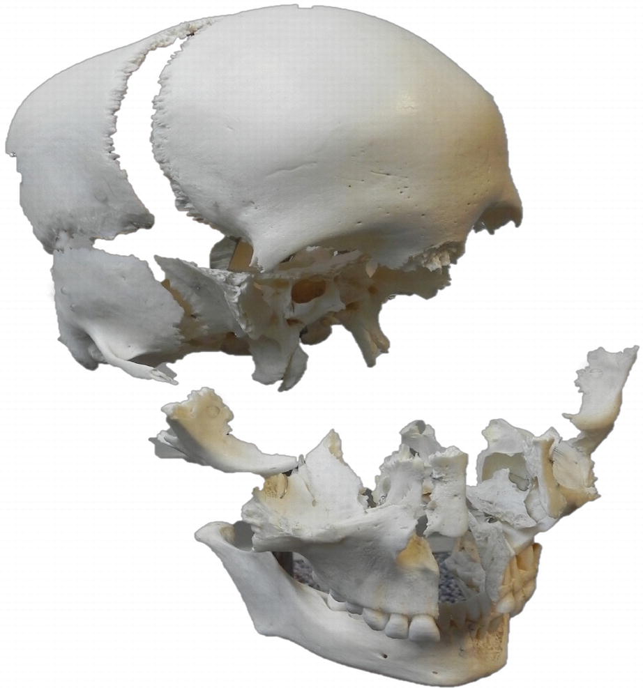
Beauchenne preparation of the human skull noting the articulations between many of the bones of the skull

Fig 12 3D reconstructed CT of the skull anterior and posterior views The - photo 4
Fig. 1.2

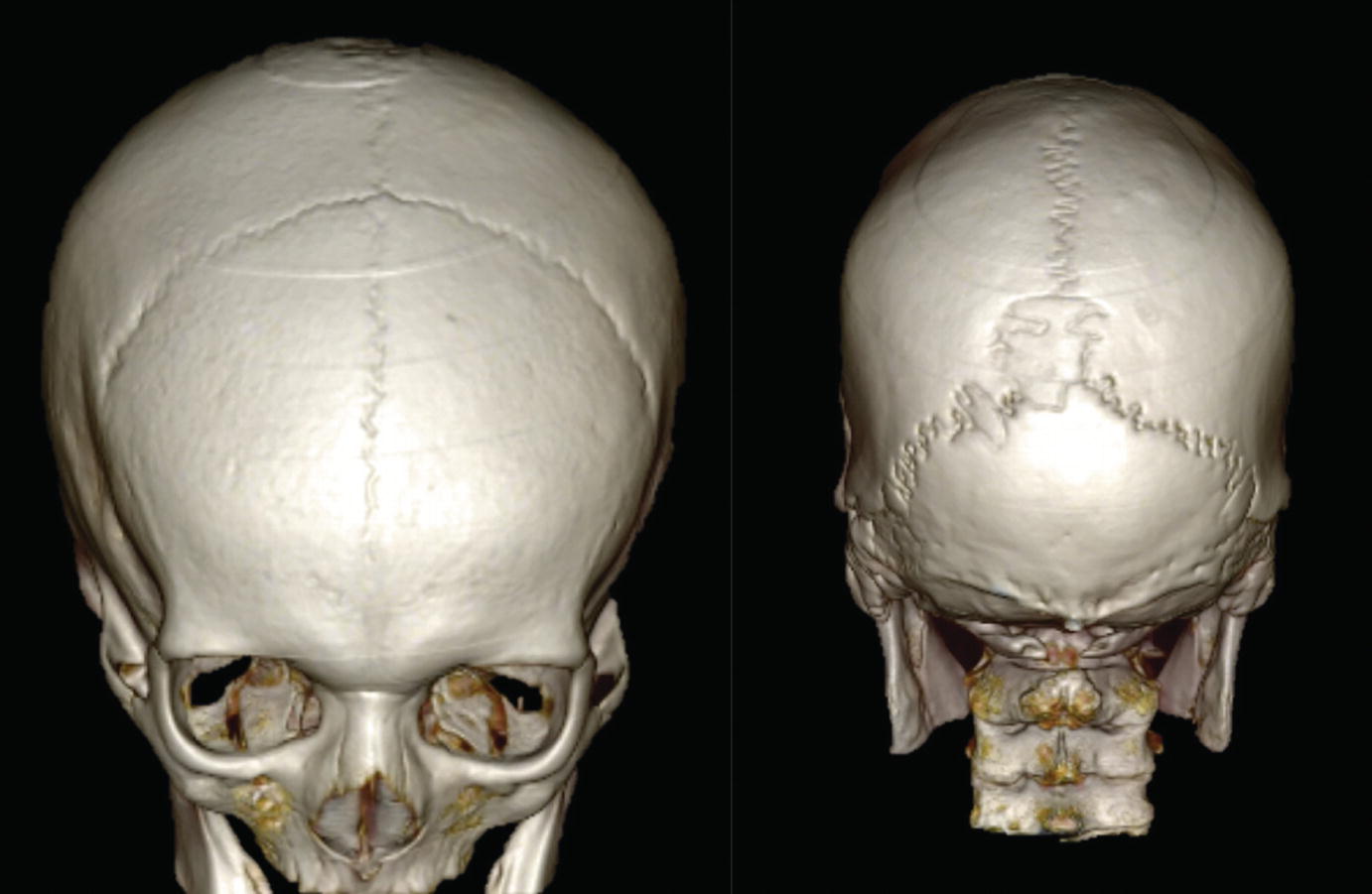
3D reconstructed CT of the skull anterior and posterior views. The anterior view (left) illustrates the sagittal, coronal and metopic sutures. The posterior view (right) notes the sagittal suture and associated sutural bone and lambdoid sutures

Fig 13 Drawings of the human skull illustrating the sutures of the calvaria - photo 5
Fig. 1.3

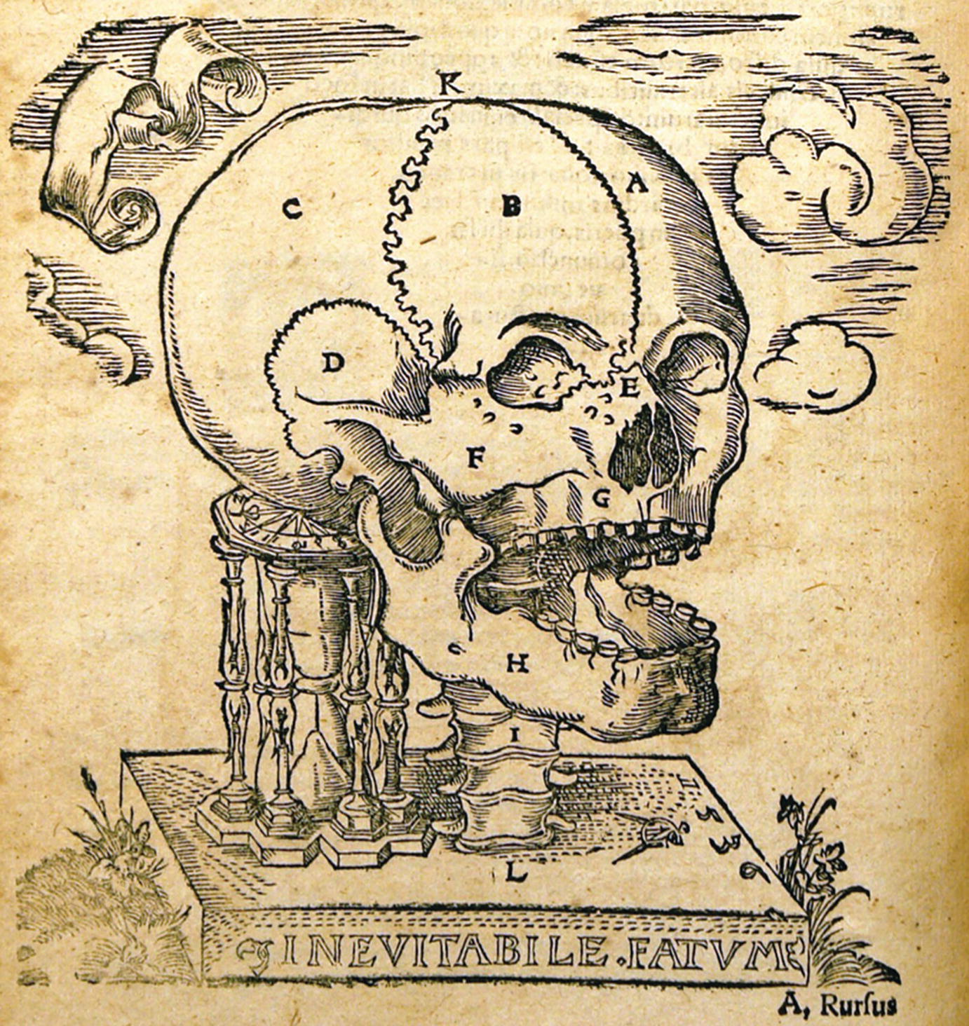
Drawings of the human skull illustrating the sutures of the calvaria from Johann Dryanders Anatomia capitis humani published in 1536

Fig 14 Drawings of the human skull illustrating the sutures of the calvaria - photo 6
Fig. 1.4

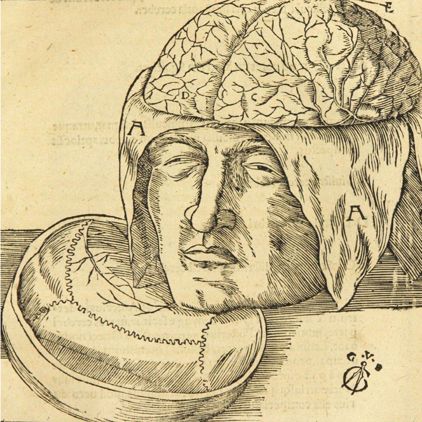
Drawings of the human skull illustrating the sutures of the calvaria from Johann DryandersAnatomia capitis humani published in 1536

Fig15 Drawing by Leonardo da Vinci 14521519 noting the coronal sutures - photo 7
Fig.1.5

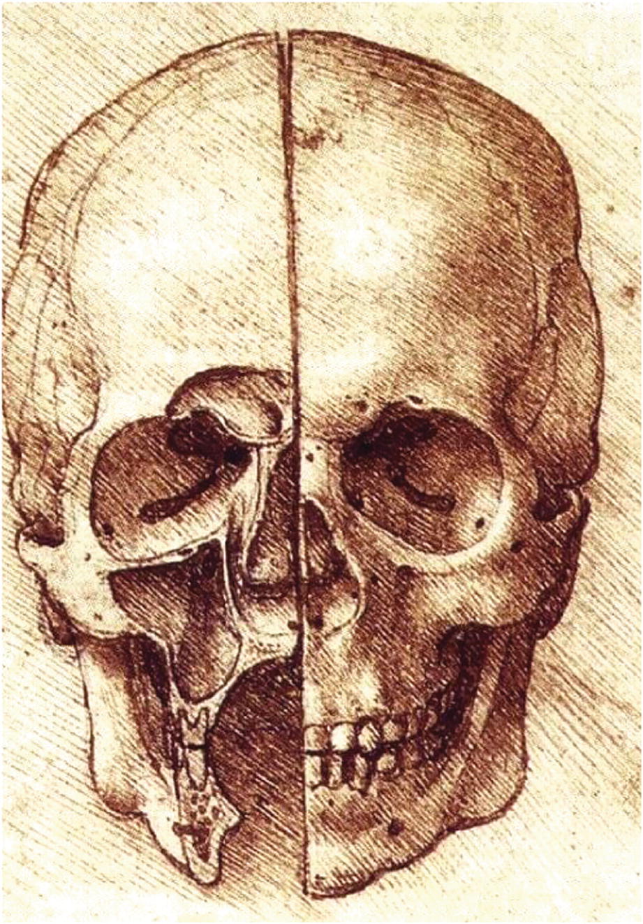
Drawing by Leonardo da Vinci (14521519) noting the coronal sutures

Fig 16 Internal view of the right half of the sagittal suture from a - photo 8
Fig. 1.6

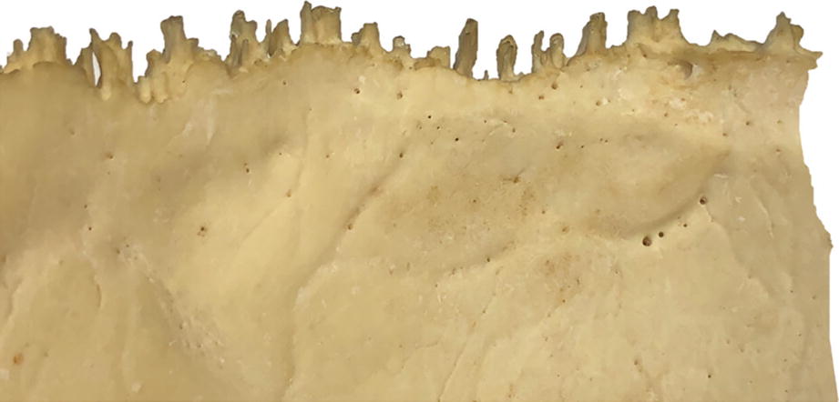
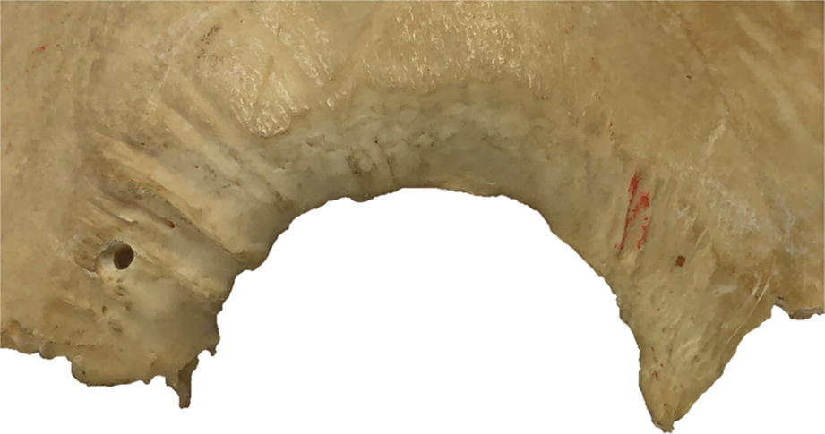
Internal view of the right half of the sagittal suture from a disarticulated parietal bone

Fig 17 External view of the right half of the sagittal suture from a - photo 9
Fig. 1.7

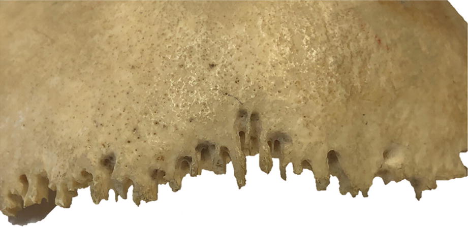
External view of the right half of the sagittal suture from a disarticulated parietal bone

Fig 18 External view of the right parietal part of the squamous suture from a - photo 10
Next page
Light

Font size:

Reset

Interval:

Bookmark:

Make

Similar books «The Sutures of the Skull: Anatomy, Embryology, Imaging, and Surgery»

Look at similar books to The Sutures of the Skull: Anatomy, Embryology, Imaging, and Surgery. We have selected literature similar in name and meaning in the hope of providing readers with more options to find new, interesting, not yet read works.


Reviews about «The Sutures of the Skull: Anatomy, Embryology, Imaging, and Surgery»

Discussion, reviews of the book The Sutures of the Skull: Anatomy, Embryology, Imaging, and Surgery and just readers' own opinions. Leave your comments, write what you think about the work, its meaning or the main characters. Specify what exactly you liked and what you didn't like, and why you think so.