Copyright 2013 Dr. Jeffrey L Williams
All rights reserved.
ISBN: 1481916602
ISBN-13: 9781481916608
eBook ISBN: 978-1-63003-284-5
Library of Congress Control Number: 2013900670
CreateSpace Independent Publishing Platform
North Charleston, South Carolina
Disclosure: The information and images included in this book are for educational purposes only. It is not intended nor implied to be professional medical advice or a substitute for professional medical advice. The reader should always consult his or her healthcare provider to determine the appropriateness of the information for their own situation or if they have any questions regarding a medical condition or treatment plan. Reading the information in this book does not create a physician-patient relationship. Trademarks, product names, or logos featured or referred to in this book are the property of the trademark or logo holders and are used for informational purposes only. Images and patient scenarios have been altered for purposes of anonymity and are used for educational purposes only.
Acknowledgements: To my wife and three great kids without whose never-failing patience (and sometimes frustration) I would have never completed this book. To my patients who tolerate my furious pace and bad jokes.
Table of Contents:

The hectic pace of todays doctors has shortchanged the information-sharing process for patients undergoing pacemaker implantation. Patients and their families are often unaware of many critical issues involved in pacemaker implantation. Though pacemaker implantation can usually be performed with minimal risks, any surgery entails risks that are particular for each procedure and patient. I have found it increasingly difficult to provide a complete consultation, physical exam, and discussion about the risks, benefits, and alternatives of pacemaker implantation in a typical forty-five-minute session. Since starting the Heart Rhythm Center in 2008, I have developed several iterations of written and online patient-education materials to complement our office discussions. This book serves as a comprehensive summary of the steps involved with pacemaker implantation: from the initial evaluation and implant procedure to the possible postoperative complications and required long-term follow-up care for patients and their caregivers (both professional and laypeople alike). Furthermore, many of my extremely elderly patients rely upon their spouses and families for help with the decision-making process and long-term care of their device. This book serves as a thorough means to ensure that all family members understand the roles, risks, and required follow-up for pacemaker patients.
Approximately 140,000 patients currently undergo pacemaker implantation in the United States each year.
This is the first and only book dedicated solely to patients, families, and their care providers as a comprehensive review of the what, why, and how of pacemaker implantation. As our patients become more and more invested in decisions that affect their health care, more detailed information is necessary so patients can make a comprehensive assessment prior to proceeding with any surgical procedure. This book can be part of the informed-consent process, because it covers more thoroughly the entire pacemaker implantation process than can be presented in a single (or even multiple) office visit(s). A particular emphasis will be placed on the complications that can occur during/after pacemaker implantation and how to assess a particular pacemaker implanters odds of a successful operation. Finally, patients concerned that they may need a pacemaker will find this book a useful summary of the complete evaluation that is performed to see if a pacemaker may be of benefit.

Basics of Heart Anatomy
and Conduction System:
Blood flow through the heart. depicts the basic structure of the heart. Blood returns from the body and enters the right atrium. The blood leaves the right atrium through the tricuspid valve, and it enters the right ventricle. The right ventricle then pumps the blood through the pulmonary valve into the lungs. The blood is oxygenated in the lungs and is returned to the left atrium. The blood leaves the left atrium through the mitral valve and enters the left ventricle. The left ventricle pumps the oxygenated blood through the aortic valve to the rest of the body; it then returns to the heart via the right atrium.
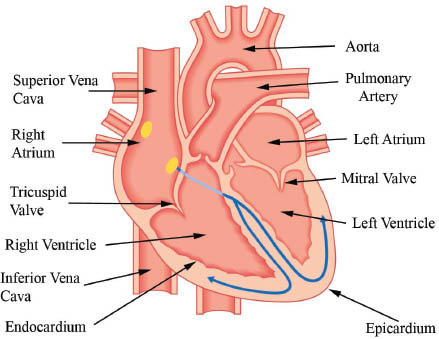
Figure 1: Basic Anatomy of the Heart
If the coronary arteries of the heart represent the plumbing, then the conduction system of the heart represents the wiring. represents the conduction system of the heart. Normal heart rate is sixty to one hundred beats per minute (bpm). Bradycardia is an abnormally slow heart rate less than 60 bpm, and tachycardia refers to an abnormally fast heart rate greater than 100 bpm. The sinoatrial (SA) node serves as the internal clockor native pacemakerof the heart and signals the appropriate heart rate for a given situation.
Electrical conduction system of the heart: The hearts natural pacemaker (the SA node) is located in the top right chamber of the heart: the right atrium. The SA node sends a signal to the upper chambers (the right and left atria) and the lower chambers (the right and left ventricles) via the atrioventricular (AV) node. The AV node transmits the electrical signal to the bottom chambers via the left and right bundle branches. The left bundle branch is comprised of left posterior and left anterior fascicles. Fascicles is another term for divisions or branches. Often, the patient has a slow heart rate because the electrical connection between the top (the signal from the SA node) and bottom (the ventricles) of the heart is diseased, called AV block. The most common type of pacemaker involves placing leads in the right atrium and right ventricle.
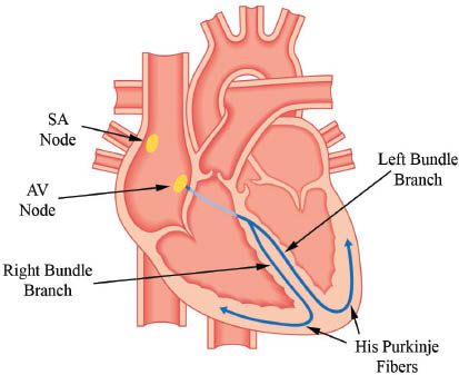
Figure 2: Conduction System of the Heart
Coronary arteries: The coronary arteries supply blood (and hence, oxygen) to the heart muscle and course along for blood supplies of various elements of the conduction system. One can see why AV Block is often seen during heart attacks (blocked coronary artery) involving the RCA, because 80% of patients have their AV node blood supply provided by the RCA. In addition, left posterior fascicular block is uncommonly due to coronary disease, because it has a dual blood supply. It would require two occluded coronary arteries (PDA and LAD septal perforators) to become blocked.
Table 1. Blood Supply to the Hearts Electrical Conduction System
STRUCTURE | BLOOD SUPPLY (% OF PATIENTS) |
SA Node | 55% RCA; 35% Left Circumflex; 10% Dual |
AV Node | 80% RCA; 10% Left Circumflex; 10% Dual |
Next page
