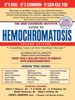
THE IRON DISORDERS INSTITUTE
GUIDE TO
HEMOCHROMATOSIS
SECOND EDITION
SCIENTIFIC ADVISORS:
Robert T. Means Jr., MD P. D. Phatak, MD E. D. Weinberg, PhD
Herbert Bonkovsky, MD Ralph G. DePalma, MD
CHERYL GARRISON, Editor Cofounder, Iron Disorders Institute

Copyright 2009 by the Iron Disorders Institute
Cover and internal design 2009 by Sourcebooks, Inc.
Cover Design by Bruce Gore/Gore Studio Design
Text design: Lisa Taylor
Sourcebooks and the colophon are registered trademarks of Sourcebooks, Inc.
All rights reserved. No part of this book may be reproduced in any form or by any electronic or mechanical means including information storage and retrieval systemsexcept in the case of brief quotations embodied in critical articles or reviewswithout permission in writing from its publisher, Sourcebooks, Inc.
This publication is designed to provide accurate and authoritative information in regard to the subject matter covered. It is sold with the understanding that the publisher is not engaged in rendering legal, accounting, or other professional service. If legal advice or other expert assistance is required, the services of a competent professional person should be sought.From a Declaration of Principles Jointly Adopted by a Committee of the American Bar Association and a Committee of Publishers and Associations
This book is not intended as a substitute for medical advice from a qualified physician. The intent of this book is to provide accurate general information in regard to the subject matter covered. If medical advice or other expert help is needed, the services of an appropriate medical professional should be sought.
All brand names and product names used in this book are trademarks, registered trademarks, or trade names of their respective holders. Sourcebooks, Inc., is not associated with any product or vendor in this book.
Published by Cumberland House, an imprint of Sourcebooks, Inc.
P.O. Box 4410, Naperville, Illinois 60567-4410
(630) 961-3900
Fax: (630) 961-2168
www.sourcebooks.com
Library of Congress Cataloging-in-Publication Data
The Iron Disorders Institute guide to hemochromatosis / [edited by] Cheryl Garrison. 2nd ed.
p. cm.
Includes bibliographical references and index.
1. HemochromatosisPopular works. I. Garrison, Cheryl D. II. Iron Disorders Institute. III. Title: Guide to hemochromatosis.
RC632.H4I76 2009
616.152dc22
2009030761
Printed and bound in the United States of America.
VP 10 9 8 7 6 5 4 3 2 1
DISCLAIMER
Content in this publication is not intended to be a substitute for professional medical advice, nor is it intended to replace the services of a medical professional or impair the relationship between patients and healthcare providers or caregivers. Content in this publication should be used as information only and be discussed with a medical professional, who is the only one qualified to diagnose and treat your condition. The Iron Disorders Institute, all contributors, and the publisher of this book are not responsible for the content on websites listed in the resources, nor do we endorse products promoted on such sites. We advise you to always seek the advice of a qualified health provider before making any changes to your diet or supplementation, or before starting any new treatment. Any application of the information or recommendations in this book is made at the readers discretion.
Foreword
Until the discovery of the HFE gene and its mutations, hereditary hemochromatosis (HHC) was not a commonly diagnosed condition. However, we now know that HHC is the leading cause of iron-overload disease and the most common hereditary disease that affects Caucasians of northern European ancestry. The common form of HHC is due to a mutation at a single point in the HFE gene. Two of these mutations (known as an autosomal recessive condition) must usually be present in a person in order for that person to develop heavy iron overload. Such people have lifelong excessive intestinal absorption of iron, which leads to deposition of the metal in the liver, heart, pancreas, and other organs. Excess iron can be toxic and even fatal. Timely diagnosis of HHC and treatment with phlebotomy can prevent organ damage. Physicians can perform a simple blood test to determine whether a patient is at risk for iron overload. This test is the serum transferrin iron-saturation percentage (TS percentage). If TS percentage is elevated above 55 percent, a repeated fasting test should be run. If the fasting TS percentage is greater than 45 percent, the physician and patient should suspect iron overload and proceed with additional tests such as serum ferritin and HFEgene mutational analysis.
Two mutations in the HFE gene have been linked to the hemochromatosis phenotype: the major mutation is called C282Y, and the minor mutation, called H63D, is less strongly associated with HHC. Iron overload occurs chiefly in people who have two copies of the C282Y mutation (homozygous, or C282Y +/+) and less often in those who have one copy of each compound, heterozygous for both C282Y and H63D (C282Y +/ or H63D /+). Tests for both are now widely and commercially available; the tests have helped to establish definitive diagnoses, particularly in patients and their families with the classic form of HHC.
In the United States, about 80 to 85 percent of patients with iron overload have the mutations in the HFE gene associated with HHC. However, many people who are homozygous for the major mutation (C282Y +/+), especially women, do not express the iron-overload phenotype. Then, too, there are other types of HHC (e.g., those occurring in black Africans or Melanesians) in which causative mutations occur in other still-unidentified genes.
Before discovery of the HFE gene and identification of the major mutations that cause HHC, physicians relied on liver biopsy for diagnosis because the liver is the principal organ responsible for the storage and detoxification of iron. It is also the first and foremost organ damaged by heavy iron overload. Indeed, the gold standard for the diagnosis of iron overload is still liver biopsy, to determine the hepatic iron index. The hepatic iron index is defined as the hepatic iron concentration, expressed as micromoles of iron per gram of dry liver, divided by the age of the patient in years. Values greater than 1.9 indicate HHC. Liver biopsy, however, is invasive and may, though rarely, be associated with complications. Furthermore, there may be considerable variability in measured hepatic iron concentrations in small liver samples from patients with cirrhosis. Genetic testing has revolutionized diagnosis of HHC because doctors can now make firm diagnoses in most patients without liver biopsies. Biopsies continue to be important to assess the presence and severity of hepatic fibrosis (scar tissue). However, among C282Y +/+ patients who have normal liver enzymes in the serum (AST, ALT) and normal livers on physical examination, when serum ferritin levels are less than 1,000 ng/mL, we now know that cirrhosis is unlikely.
We also have made progress in developing noninvasive methods for estimating hepatic iron concentrations. A dedicated device for measuring magnetic susceptibilities, the superconducting quantum interference device, is accurate for the estimation of hepatic iron concentrations but is not widely available. Although computed tomography (CT) scanning demonstrates an increase in the attenuation of the liver in hepatic iron overload, it is relatively insensitive to mild degrees of increased hepatic iron, especially if there is associated fatty change in the liver. Special magnetic resonance imaging (MRI) is a more promising noninvasive modality for estimating hepatic iron concentration. An increase in hepatic iron concentration decreases the signal intensity and makes the liver appear dark on typical MR images. Some centers now use a test called FerriScan, which uses special MRI algorithms to provide an accurate estimate of hepatic iron concentrations.


