THE RESPIRATORY SYSTEM
THE HUMAN BODY
THE
RESPIRATORY
SYSTEM
EDITED BY KARA ROGERS, SENIOR EDITOR, BIOMEDICAL SCIENCES

Published in 2011 by Britannica Educational Publishing
(a trademark of Encyclopdia Britannica, Inc.)
in association with Rosen Educational Services, LLC
29 East 21st Street, New York, NY 10010.
Copyright 2011 Encyclopdia Britannica, Inc. Britannica, Encyclopdia Britannica, and the Thistle logo are registered trademarks of Encyclopdia Britannica, Inc. All rights reserved.
Rosen Educational Services materials copyright 2011 Rosen Educational Services, LLC. All rights reserved.
Distributed exclusively by Rosen Educational Services.
For a listing of additional Britannica Educational Publishing titles, call toll free (800) 237-9932.
First Edition
Britannica Educational Publishing
Michael I. Levy: Executive Editor
J.E. Luebering: Senior Manager
Marilyn L. Barton: Senior Coordinator, Production Control
Steven Bosco: Director, Editorial Technologies
Lisa S. Braucher: Senior Producer and Data Editor
Yvette Charboneau: Senior Copy Editor
Kathy Nakamura: Manager, Media Acquisition
Kara Rogers: Senior Editor, Biomedical Sciences
Rosen Educational Services
Heather M. Moore Niver: Editor
Nelson S: Art Director
Cindy Reiman: Photography Manager
Matthew Cauli: Designer, Cover Design
Introduction by Amy Miller
Library of Congress Cataloging-in-Publication Data
The respiratory system / edited by Kara Rogers.
p. cm. -- (The human body)
In association with Britannica Educational Publishing, Rosen Educational Services.
Includes bibliographical references and index.
ISBN 978-1-61530-254-3 (eBook)
1. Respiratory organsPopular works. I. Rogers, Kara.
QP121.R467 2011
612.2dc22
2010014243
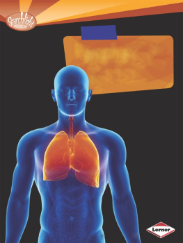
On the cover: The human lungs are extraordinary organs that constantly pump crucial oxygen through airways and into the bloodstream. www.istockphoto.com / Sebastian Kaulitzki
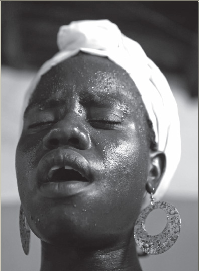
On page : Singing is one of many common activities that requires dynamic breath control. Chip Somodevilla/Getty Images
On pages : A healthy set of lungs is the powerhouse behind the respiratory system. www.istockphoto.com / nicoolay
CONTENTS



INTRODUCTION
T he human lungs are amazing feats of nature. They pump vital oxygen through airways and into the bloodstream every second of every day. Without this ability, humans could not survive on Earth.
This book explains the science behind the amazing human respiratory system. It also sheds light on how easily a healthy respiratory system can be damaged, whether by a viral or bacterial infection or through detrimental habits such as smoking. But there are many treatments to keep the airways free and clear, and this book also describes the many different approaches doctors can take to save patients lives and lungs.
The anatomy of the human respiratory system starts at the place where air first enters the bodythe nose. This structure provides humans with the sense of smell while also filtering, warming, and moistening inhaled air.
Air that passes through the nose travels to the pharynx, or throat, the cone-shaped passageway leading from the mouth and nose to the larynx, or voice box. The larynx is a hollow tube connected to the top of the windpipe, and this air canal to the lungs not only enables humans to speak but also keeps food out of the lower respiratory tract.
After passing through the larynx, air travels through the trachea, also known as the windpipe. Here, the air is cleansed and moistened before entering the lungs. The clean air then travels into the deep tissues of the lungs, eventually reaching the region where gas is exchanged, the centre of the respiratory system.
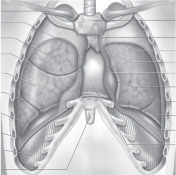
The right lung is slightly larger than the left lung because of the asymmetrical position of the heart. The right lung has 10 airway segments, and the left lung has 8 to 10. A thin membranous sac known as the pleura covers the lungs. Inside the lungs, there are numerous nerves and blood vessels. However, the most prominent feature of the lung interior are the many small air passages called bronchioles, which range in diameter from 3 mm (0.12 inch) to less than 1 mm (less than 0.04 inch).
The gas-exchange area, the region where oxygen is transferred to the blood and carbon dioxide is removed, is made up of three separate compartments for blood, air, and tissue. The tissue compartment supports the air and blood compartments and lets them come into close contact, which makes exchanging gases easier, but still keeps them separate. The exchange of carbon dioxide and oxygen takes place in tiny air sacs called alveoli, which look like cells in a honeycomb. The average adult lung has approximately 300 million alveoli.
Lungs also have two distinct blood circulation systems. The first of these, the pulmonary system, is characterized by the transport of carbon dioxideladen blood from the right side of the heart, through the pulmonary arteries, and to the lungs and by the subsequent transport of oxygen-rich blood from the lungs, through the pulmonary veins, and to the left atrium of the heart. From the heart, the oxygenated blood is pumped to the rest of the body, thereby delivering oxygen and other nutrients to organs distant from the lungs. The second blood system in the lungs, the bronchial circulation, comprises the network of blood vessels supporting the conducting airways themselves. The bronchial circulation is a vital source of nourishment for the lung tissues.
The act of breathing, or respiration, is an automatic process, controlled by the brain. Thus, humans and other animals do not need to actively think about breathing in order for it to happen. A significant feature of the human respiratory system is its capacity to instantly adjust to internal and external stimuli on its own. A series of neural networks in the brain control the rate of breathing by communicating with the muscles in the chest and the abdomen. One of the major abdominal muscles involved in breathing is the diaphragm, which functions to move air in and out of the lungs as it contracts and relaxes, respectively.
Next page
