Normal BBB Structure and Function.
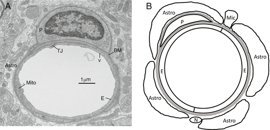
In contrast to systemic capillaries, brain capillary endothelial cells are linked by TJs and have a low basal rate of transcytosis (Fig. ]).
Fig. 1
( a ) An electron micrograph showing the structure of a normal mouse cerebral capillary. ( b ) A schematic showing the relationship between the endothelium and other elements of the neurovascular unit. Brain capillary endothelial cells (E) are linked by tight junctions (TJ), show few vesicles (v) and have more mitochondria (mito) than systemic capillaries. Sharing the same basement membrane (bm) as the endothelium are pericytes (P). Capillaries are surrounded by astrocyte endfeet (astro), with occasional neuronal (N) and microglial processes (Mic). The endothelial cells and the ensheathing cells/basement membrane are called the neurovascular unit
Endothelial TJs are integral for BBB function. They are comprised of transmembrane proteins, which form the links between adjacent endothelial cells, and cytoplasmic plaque proteins that form a physical scaffold for the TJs and regulate TJ function [].
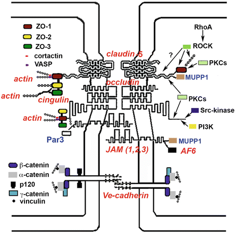
Fig. 2
Schematic representation of the basic components of the brain endothelial junctional complex. The left of the panel shows potential structural components of the TJ complex. Claudin-5, occludin, and the JAMs are transmembrane proteins that link adjacent cells. Claudin-5 is the major occlusive protein at the BBB while the cytoplasmic plaque protein, ZO-1, is important for clustering of claudin-5 and occludin and establishing connection with actin filaments. The roles of ZO-2, ZO-3, MUPP1, Par3, Af6 are less clear in brain endothelial cells. Brain endothelial cells also possess adherens junctions, containing the transmembrane protein, Ve-cadherin, and cytosolic accessory proteins (e.g., catenins). The right of the panel shows signaling molecules involved in modulating TJ complex assembly and disassembly
Transmembrane proteins : These include the claudins, occludin, and the junctional associated molecules (JAMs). In addition, tricellulin, a molecule with homology to occludin, is present at points of three cell contact. The transmembrane proteins can form trans -interactions with proteins on other cells or cis -interactions within the same plasma membrane [].
The claudins are a 27 gene family which are involved in closing the paracellular space between cells (e.g., claudin-5) and in forming ion pores (e.g., claudin-2) [].
Although occludin is not directly involved limiting paracellular permeability, it is thought to be a central regulatory component of the TJ being involved in TJ formation and maintenance [].
Junctional adhesion molecules (JAM-A, -B, -C) are members of the immunoglobulin superfamily. Their role at the BBB has received much less attention than claudin-5 and occludin, but evidence indicates they are involved in regulating not only paracellular permeability but also leukocyte/endothelial interactions [].
Cytoplasmic plaque proteins : As well as the transmembrane proteins, there are an array of cytosolic proteins associated with the TJs that form the cytoplasmic plaque (Fig. ]), they can act as scaffolding proteins linking different elements of the TJ (e.g., transmembrane proteins, cytoplasmic plaque proteins, and the actin cytoskeleton).
Cytoplasmic plaque proteins that do not contain PDZ domains include cingulin, 7H6, Rab13, ZONAB, AP-1, PKC, and PKC, as well as several G proteins (Gi, Gs, G12, Go). These proteins have multiple functions. Thus, for example, it is thought that cingulin acts as a cross link between TJ transmembrane proteins and the actin-myosin cytoskeleton and that the PKC isoforms and the G proteins regulate TJ assembly and maintenance [].
Adherens junctions : Cerebral endothelial cells possess adherens junctions (AdJs) as well TJs. AdJs are also a complex of transmembrane (Ve-cadherin) and cytosolic accessory proteins (, catenin, p120) closely associated with actin filaments [].
Cell cytoskeleton : The cytoskeleton comprises of actin microfilaments, intermediate filaments, and microtubules. The actin cytoskeleton is linked to the TJs via cytoplasmic plaque proteins such as ZO-1 and to AdJs via catenins (Fig. ]. Changes in that cytoskeleton may impact TJ function by altering the physical support (scaffold) for the junction and by transmitting physical forces to the junction proteins.
Neurovascular unit (NVU): Although the cerebral endothelial cells and their linking TJs are the ultimate determinants of BBB permeability, perivascular cells (pericytes, astrocytes, neurons, microglia) have a large role in regulating that permeability. In particular, pericytes, which share the same basement membrane as the endothelium, and astrocytes, whose endfeet almost completely surrounds cerebral capillaries (Fig. ]. These cells, together with smooth muscle cells in larger vessels, act in concert to regulate cerebrovascular function (blood flow and barrier function), forming the NVU.

