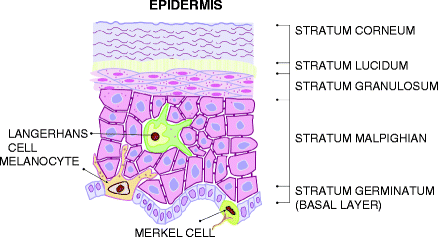
The epidermis consists of many cells, about 95% are keratinocytes, and the other prominent cells are melanocytes, Langerhans cells, and Merkels cells. The epidermis does not have any blood vessels; it obtains its nutrients from the blood vessels of the dermis diffusing through the dermoepidermal junction (Fig. ).
Figure 1.1.
Epidermal layers: stratum corneum - anucleated cells; stratum lucidum - present only in palms and soles; stratum granulosum - epidermal nuclei start disintegrating; stratum malpighian - thickest and strongest layer; stratum germinatum - the only cells which undergo division. Epidermal cells: keratinocytes - the main cells of the epidermis, present in every layer of the epidermis; melanocytes - dendritic pigment producing cells, seen with a halo around them under ordinary staining, due to the lack of desmosomes. Present amongst the basal cells; langerhans cells - dendritic immunologically competent cells, also seen with a halo around them, due to the absence of desmosomes. Present in the stratum malpighian; merkel cells - present only in hairless skin; related to the sense of touch. These cells can only be seen under an electron microscope. Present amongst the basal cells
1.1.1.1 Keratinocytes
The main function of keratinocytes is to produce keratin. Keratin forms the internal skeleton of the keratinocytes, and is composed of intermediate filaments. The keratins are a family of 30 proteins each produced by a different gene; different keratins are found in different levels of the epidermis, depending upon the stage of differentiation.
Keratinocytes are arranged in layers. The innermost layer is the basal layer; changes occur in the cells as they move up to produce the tough, impermeable, fibrous protein keratin present in the outermost layer, the stratum corneum. The cells get larger, thinner, and flatter as they move upwards.
Basal Cell Layer (Stratum Germinatum)
These are the innermost cells, present as a single layer over the basement membrane. These are the only cells of the epidermis that divide. The cells are columnar in shape with large, dark-staining nuclei. The basic component of the cytoplasm is the tonofilament - the keratin filament.
The cell cycle time is the time between two successive episodes of mitosis, or the time taken for the individual cells to divide. The normal cell cycle time is 163 h; in psoriasis it is reduced to 37.5 h.
Stratum Malpighian (Stratum Spinosum)
This layer consists of a number of layers (four to ten) of polyhedral cells. The cells have a central oval nucleus; the cytoplasm is packed with tonofilaments. Intercellular bridges called desmosomes connect the cells to one another. The cells also have a number of organelles, which help in the formation of keratin and intercellular adhesion of the stratum corneum.
Stratum Granulosum
The layer is so called because of the granules it contains. These granules (keratohyaline granules) contain proteins that help in the aggregation of the keratin filaments. It also consists of proteins that help to bind the cells of the stratum corneum together. It is three to four layers in thickness; the cells are diamond shaped.
Stratum Lucidum (Only in the Palms and Soles)
This layer lies between the stratum granulosum and stratum corneum; it is only present in the palms and soles, where the skin is very thick. The cells are nucleated, have opaque membranes and dense cytoplasm.
Stratum Corneum
An abrupt transition occurs as the cells move up from the stratum granulosum to the stratum corneum. The viable nucleated cells of the stratum granulosum change to anucleated dead cells of the stratum corneum. The cells of the stratum corneum are large, flat polyhedral, and filled with keratin; they vary in thickness from 15 to 25 layers. The cells are held together by firm lipid-rich cement. The cells overlap each other; this further helps in making this layer more impenetrable.
The upper layers of the stratum corneum are shed from the skin surface in the form of microscopic scales. The same number of cells lost from the surface are replaced by the cells of the basal layer. It takes about 26-42 days for the cells to migrate from the basal layer to the top of the granular layer, and another 13-14 days for the cells to cross the stratum corneum to the surface, from where they are shed. Total transit time is 52-75 days. In psoriasis, it is reduced to 8-10 days.
1.1.1.2 Melanocytes
These are the pigment producing cells of the epidermis; they are derived from the neural crest. They are present in the basal layer. The number varies in different parts of the body; on an average there is one melanocyte to every ten basal cells. Melanocytes are dendritic cells; they transfer melanin through the dendrites to the keratinocytes to protect them from ultraviolet light and to give color to the skin. Melanin granules form a protective cap over the outer part of the keratinocyte in the inner layers of the epidermis. In the stratum corneum, they are uniformly distributed to form a UV-absorbing blanket, which reduces the amount of radiation penetrating the skin. The melanin is present within granules called melanosomes. Melanin is formed from the aminoacid tyrosine with the help of the enzyme tyrosinase.
The number of melanocytes is the same in every individual; it is the size of the melanosomes and the distribution of melanin, which is responsible for the complexion of their skin.
1.1.1.4 Merkel Cells
These cells are also found in the basal cell layer, in close proximity to the hair follicles. They act as transducers of fine touch. Their cytoplasm contains neuropeptide granules, as well as neurofilaments and keratin.