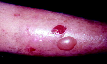
Following her hospitalization, she was moved to her daughter-in-laws home. After 1 month, the daughter-in-law noticed malodor emanating from the patients legs, raising the possibility of superinfection. The patient also complained of pruritus and pain. She was hospitalized, the bandages were removed, and multiple tense serous blisters were noted from the mid-calf to the feet.
The patient had temporary symptom relief with oral steroids but quickly had recurrence after steroid taper was attempted three times. The patient noted discoloration, thinning and dryness of her skin.
Biopsies for direct immunofluorescent studies were obtained from perilesional skin on the right shin and right buttocks and were positive for linear junctional IgG and C3.
Discussion
Bullous pemphigoid, which is caused by autoantibodies that are directed against proteins at the dermal-epidermal junction, is an immunoblistering disease (Abbas et al., ).
This disease generally affects the elderly. The lesions appear as tense, fluid filled bullae. They can be pruritic and associated with erythematous or normal skin, but sometimes can have an urticarial appearance. Lesion distribution is often the inner thighs, flexor surfaces of the forearms, axillae, groin, lower legs and lower abdomen. The lower legs are often the first site of manifestation. Bullous pemphigoid has also been linked to other disorders, such as diabetes, rheumatoid arthritis, dermatomyositis, ulcerative colitis, myasthenia gravis, and thymomas (Bolognia ). Many cases of bullous pemphigoid enter into remission within a few years of diagnosis.
Pemphigus vulgaris is also an autoimmune disease. In contrast to bullous pemphigoid, antidesmoglein antibodies are directed against the intercellular surfaces of epidermal keratinocytes (Bolognia ). Thus, cell-cell adhesion is disrupted, bullae break apart easily, and shallow erosions covered with crust are the usual clinical findings. Unlike bullous pemphigoid, pemphigus vulgaris is found in a younger population (patients usually in their thirties or forties). Lesions in pemphigus vulgaris usually start in the oral mucosa and then spread to the skin. Pemphigus vulgaris can involve the scalp, face, axilla, groin, trunk and points of pressure. These lesions are painful and less commonly itch. Nikolsky sign is also present, with easy skin removal by applying a slight shearing force on normal-appearing skin adjacent to the lesion.
Epidermolysis bullosa acquisita (EBA) is worth mentioning in the differential diagnosis of this patient because it shares some clinical and histological features of bullous pemphigoid. EBA is a chronic autoimmune disease characterized by severe subepidermal blistering, scars, and milia with a tendency to form in traumatized areas of the body. A specialized form of direct immunofluorescence on salt-split skin is required to differentiate between EBA and BP (Bolognia ).
Bullous scabies can mimic the histology and direct immunofluorescent findings of bullous pemphigoid and therefore must be included in the differential diagnosis (Shahab ). History and mineral oil mounted skin scrapings can inexpensively and rapidly make the appropriate diagnosis.
Based on the patients medical history, clinical picture, and biopsy results, the diagnosis of bullous pemphigoid was made. Of note, direct immunofluorescent skin biopsies should be obtained from perilesional skin rather than intact blisters or erosions to have the best diagnostic sensitivity.
The patient was instructed to avoid sun exposure and keep the lesions dry and clean. Topical clobetasol cream and tetracycline 500 mg bid were prescribed with good results. If a flare occurs systemic immunosuppressives could be considered as a next step.
References
Abbas AK, Fausto N, Kumar V. Robbins and Cotran pathologic basis of disease. 7th ed. Philadelphia: The Curtis Center; 2005. p. 12602.
Bolognia J, Jorizzo JL, Rapini RP. Dermatology. St. Louis: Mosby/Elsevier; 2008.
Johnson RA, Suurmond D, Wolff K. Fitzpatricks color atlas & synopsis of clinical dermatology. 5th ed. New York: McGraw-Hill; 2005. p. 1008.
Shahab RK, Loo DS. Bullous Scabies. J Am Acad Dermatol. 2003 Aug; 49(2):346350. PubMed CrossRef

