Part I
The Core Needle Biopsy Program
Springer International Publishing Switzerland 2016
Sandra J. Shin (ed.) A Comprehensive Guide to Core Needle Biopsies of the Breast 10.1007/978-3-319-26291-8_1
1. Essential Components of a Successful Breast Core Needle Biopsy Program: Imaging Modalities, Sampling Techniques, Specimen Processing, Radiologic/Pathologic Correlation, and Appropriate Follow-Up
Christine M. Denison 1
(1)
Department of Radiology, Brigham and Womens Hospital, Harvard Medical School, Boston, MA, USA
(2)
Department of Pathology, Brigham and Womens Hospital, Harvard Medical School, Boston, MA, USA
Christine M. Denison
Email:
Susan C. Lester (Corresponding author)
Email:
Keywords
Breast core needle biopsies Quality assurance Best practices Radiologic/pathologic correlation Artifacts Core needle biopsy recurrence Correlation conference Pitfalls Specimen processing
Introduction
The ability to accurately sample and diagnose breast lesions without the need for surgery has major advantages for patients, radiologists, pathologists, surgeons, biomedical research, and the healthcare system:
Benefits to Patients
The amount of tissue needed for diagnosis is minimized.
The morbidity of a needle biopsy is less than that of a surgical excision.
Fewer biopsy-related changes (e.g., scarring) that could complicate imaging follow-up are created.
A definitive diagnosis is obtained in a short span of time.
Women with benign findings are spared surgery.
A core needle biopsy of malignancy aids in subsequent treatment planning.
Multiple areas in both breasts can be sampled to determine optimal surgery.
Patients who proceed initially with surgery are more likely to achieve the definitive excision of the breast cancer, with negative margins, and nodal evaluation in one procedure.
Patients have the option to undergo treatment first (i.e., presurgical or neoadjuvant therapy). The clip placed at the time of the needle biopsy is essential to identify the tumor bed after treatment.
Core needle biopsies obtained without the use of heating or freezing tissue obtain cancer samples with the optimal preservation of biomolecules for assays to determine therapy and prognosis.
Women with lymphoma or metastatic malignancies are spared unnecessary surgery.
Benefits to Radiologists
Lesions identified by imaging can be directly sampled to obtain optimal correlation with pathologic diagnosis.
A specific diagnosis can aid in following patients with focal findings on breast imaging over time.
Close correlation of radiologic findings with pathologic diagnoses increases the knowledge base of radiologists.
Benefits to Pathologists
Smaller core needle specimens are easier to process and evaluate compared to excisional biopsies.
There is greater certainty that the tissue being examined by the pathologist contains the lesion detected by imaging. In contrast, small lesions can be very difficult to identify with certainty in large excisional specimens and mastectomies.
Close correlation of radiologic findings with pathologic diagnoses increases the knowledge base of pathologists.
Benefits to Surgeons
Surgical planning is facilitated by the ability to evaluate multiple lesions by core needle biopsy.
It is more likely that surgery of the breast and lymph nodes will be successful in one procedure when there is a definitive diagnosis of cancer on core needle biopsy.
A clip placed at the time of diagnosis aids in localization of the tumor bed for breast-conserving therapy after neoadjuvant therapy.
Benefits for Biomedical Research
A needle biopsy provides tissue with optimal preservation of biomolecules due to minimal ischemic time and the absence of damage due to heating or freezing of the tissueif the needle biopsy technique used only cutting needles for tissue removal.
A definitive diagnosis of invasive carcinoma with determination of tumor markers on core needle biopsy provides sufficient information to evaluate patients prior to neoadjuvant therapy.
Tumors can be sampled at multiple time points during treatment.
Benefits to the Medical Care System
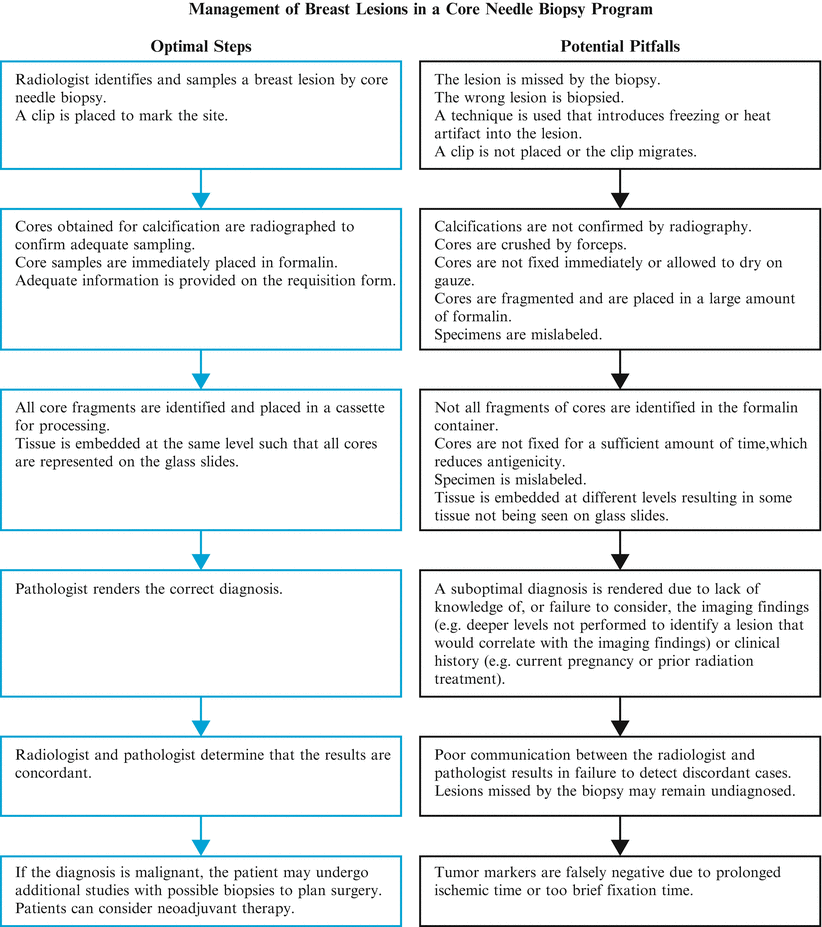
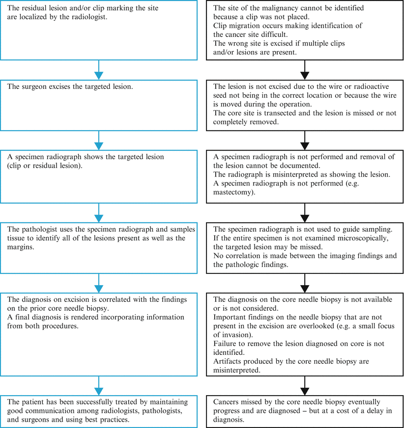
A successful core needle biopsy program requires close communication among radiologists, pathologists, and surgeons and a basic understanding of the principles of all three disciplines by everyone involved [). In this chapter the information that pathologists need to know about breast imaging, biopsy processing, radiologic/pathologic correlation, and the surgical excision of lesions identified by core needle biopsy will be discussed, as well as the critical role of the pathologist in a core needle biopsy program.
Fig. 1.1
The numerous steps and multiple people involved in a core needle biopsy program ( left side ) creates the possibility for many types of errors ( right side ). Good communication as well as an understanding of the management issues in all three disciplines (pathology, radiology, and surgery), ensures that patients have an optimal outcome
Breast Imaging Modalities
The three modalities commonly used to evaluate the breast are mammography, ultrasound, and magnetic resonance imaging (MRI).
The American College of Radiologists has created standardized terminology for reporting imaging findings (the Breast Imaging Reporting and Data System or BI-RADS) (Table ]. The majority of lesions undergoing biopsy will be in the BI-RADS 35 categories.
Table 1.1
BI-RADS assessment categories
Assessment category | Clinical management | Likelihood of cancer |
|---|
0: Incomplete | Additional imaging or comparison to prior images | Not applicable |
1: Negative | Routine screening | ~0% |
2: Benign | Routine screening | ~0% |
3: Probably benign | Short-interval follow-up or continued surveillance | >0% but 2% |
4: Suspicious | Biopsy | >2% but <95% |
4A: Low suspicion | >2% but 10% |
4B: Moderate suspicion | >10% but 50% |
4C: High suspicion | >50% but <95% |
5: Highly suggestive of malignancy |

