Published in 2015 by Britannica Educational Publishing (a trademark of Encyclopdia Britannica, Inc.) in association with The Rosen Publishing Group, Inc.
29 East 21st Street, New York, NY 10010
Copyright 2015 The Rosen Publishing Group, Inc., and Encyclopdia Britannica, Inc. Encyclopdia Britannica, Britannica, and the Thistle logo are registered trademarks of Encyclopdia Britannica, Inc. All rights reserved.
Distributed exclusively by Rosen Publishing.
To see additional Britannica Educational Publishing titles, go to rosenpublishing.com.
First Edition
Additional content and editorial input provided by Melissa Petruzzello.
Britannica Educational Publishing
J. E. Luebering: Director, Core Reference Group
Anthony L. Green: Editor, Comptons by Britannica
Rosen Publishing
Hope Lourie Killcoyne: Executive Editor
Nelson S: Art Director
Brian Garvey: Designer
Cindy Reiman: Photography Manager
Library of Congress Cataloging-in-Publication Data
Tucker, Clementine, author.
How cell processes are regulated/Clementine Tucker and Lois Sakany.
pages cm.(The Britannica guide to cell biology)
Includes bibliographical references and index.
ISBN 978-1-6227-5808-1 (eBook)
1. CytologyJuvenile literature. 2. Cell physiologyJuvenile literature. I. Sakany, Lois, author. II. Title.
QH582.5.T825 2015
571.6dc23
2014023036
Photo credits:
Cover (cell illustration) Mopic/Shutterstock.com; cover (background), pp. 1, 3, 4, 5 Sebastian Tomus/Shutterstock.com.
CONTENTS
C ells were first described in 1665 by the English scientist Robert Hooke, who published a book about his findings. Using a microscope of his own design to observe thinly sliced sections of cork, Hooke was able to see the minute, boxlike units of which the cork was composed. Hooke called these structures cells because of their similarity to the monastery cells used by monks. The first description of living cells was provided in 1674 by Dutch scientist Antonie van Leeuwenhoek, who observed bacteria and protozoa under his microscope. Ten years later, Leeuwenhoek also gave the first accurate description of red blood cells.
In the 19th century, improvements in microscopes allowed more detailed investigations of cells and their structures. In the 1830s, Scottish botanist Robert Brown discovered the cell nucleus, and two German scientists, Mathias J. Schleiden and Theodor Schwann, concluded independently that cells were the basis of all life, a view known as cell theory. Rudolf Virchow, another German scientist, stated in 1858 that all cells develop from previously existing cells. During the late 19th century, techniques of fixing and staining tissues to preserve cells opened the way for intensive research.

Scientist Robert Hooke was the first person to state that all matter expands when heated and that air is made up of particles separated from each other by relatively large distances. Private Collection/Bridgeman Images
Modern scientists, such as microbiologists and cell biologists, use a variety of microscopes to study cells. An optical, or light, microscope uses a beam of light and optical lenses for viewing a specimen. A bright-field optical microscope, in which the background is brightly lit, can achieve a power of magnification of about 1,000x. Electron microscopes, which were first developed in the early 20th century, use magnetic fields to control a beam of electrons (negatively charged particles) to get an image. Electron microscopes can produce an image with a power of magnification of up to two million times, allowing biologists to examine the structure and contents of cells at an extremely fine scale.
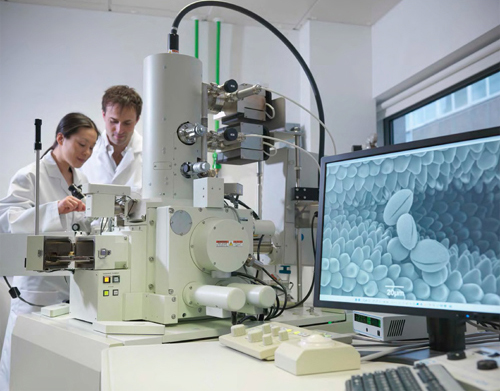
Scientists in a biolab use a scanning electron microscope to examine pollen. The microscopic image of the pollen sample appears on the monitor to the right. Monty Rakusen/Cultura/Getty Images
A cell is the smallest unit of living matter that can exist by itself. While some organisms, such as bacteria, consist of only a single cell, others, such as humans and plants, are composed of many billions of cells. Cells exist in a variety of shapes and sizes. The red blood cells in animals are disk-shaped, while many plant cells resemble cubes. While some cells, such as those found in bird eggs, can be as large as a tennis ball, most cells are microscopic. Some bacteria cells are so small that thousands could fit on the period at the end of this sentence. Given these minute sizes, cells are usually measured in microns, also known as micrometers. A micrometer (m) is equal to about one-millionth of a meter (about 25,000 micrometers equal 1 inch). The smallest bacteria are about 0.2 m in diameter. The diameter of the average human cell is roughly 10 microns, making it invisible to the naked eye.
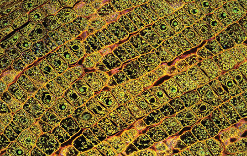
A special optical imaging technique that increases optical resolution and contrast by eliminating out-of-focus light was used to reconstruct the 3D structures of these microscopic plant cells. Fernan Federici/Moment/Getty Images
Regardless of size, every cell contains the components needed to maintain life. In multicellular organisms, the various types of cells function with great efficiency, diversifying into different parts of the organism to work together harmoniously. Even single-celled organisms, such as bacteria or some protozoans, are able to perform all of the functions necessary to their survival.
But how do cells regulate themselves? How do they know how or when to divide, where they should go, or what they should become? The book will explain how cells regulate themselves: their processes, their life cycle, and their ultimate end.
B ased on fundamental differences in their cell structure, all organisms are classified as either being prokaryotic or eukaryotic. Both prokaryotic and eukaryotic cell types contain double-stranded DNA (deoxyribonucleic acid) as their genetic material. However, eukaryotic DNA is enclosed within the nucleus while prokaryotic DNA floats freely inside the cell or sometimes forms a circular plasmid. Prokaryotic cells do not have a true nucleus and do not have structures called organelles. Prokaryotic organisms, such as archaea and bacteria, are usually single-celled and are believed to have evolved before eukaryotes.
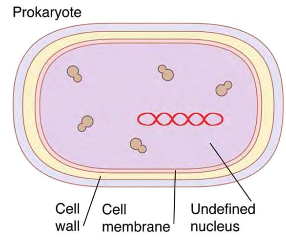
A prokaryotic cell lacks a true nucleus. Instead, nuclear matter floats freely within the cells interior. Peter Gardiner/Science Source
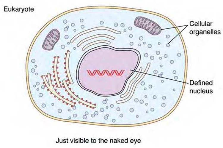
In contrast, eukaryotic cells have distinct and specialized organelles, including a nucleus to house their DNA. These membrane-bound little organs perform a variety of functions. Eukaryotic cells compose organisms ranging in size from single-celled paramecia and amoebas to all multicellular fungi, plants, and animals.
