Scientific Imaging with Photoshop
Methods, Measurement, and Output
Jerry Sedgewick

Scientific Imaging with Photoshop: Methods, Measurement, and Output
Jerry Sedgewick
New Riders
1249 Eighth Street
Berkeley, CA 94710
510/524-2178
510/524-2221 (fax)
Find us on the World Wide Web at: www.newriders.com
To report errors, please send a note to
New Riders is an imprint of Peachpit, a division of Pearson Education
Copyright 2008 by Gerald Sedgewick
Project Editor: Victor Gavenda
Production Editor: Hilal Sala
Development Editor: Anne Marie Walker
Copyeditor: Anne Marie Walker
Technical Editors: Dr. Marna Ericson and Traci Bernatchy
Proofreader: Liz Welch
Compositor: Kim Scott, Bumpy Design
Indexer: Ann Rogers
Cover design: Mimi Heft
Interior design: Mimi Heft
Notice of Rights
All rights reserved. No part of this book may be reproduced or transmitted in any form by any means, electronic, mechanical, photocopying, recording, or otherwise, without the prior written permission of the publisher. For information on getting permission for reprints and excerpts, contact .
Notice of Liability
The information in this book is distributed on an As Is basis without warranty. While every precaution has been taken in the preparation of the book, neither the author nor Peachpit shall have any liability to any person or entity with respect to any loss or damage caused or alleged to be caused directly or indirectly by the instructions contained in this book or by the computer software and hardware products described in it.
Trademarks
Adobe and Photoshop are registered trademarks of Adobe Systems Incorporated in the United States and/or other countries. Many of the designations used by manufacturers and sellers to distinguish their products are claimed as trademarks. Where those designations appear in this book, and Peachpit was aware of a trademark claim, the designations appear as requested by the owner of the trademark. All other product names and services identified throughout this book are used in editorial fashion only and for the benefit of such companies with no intention of infringement of the trademark. No such use, or the use of any trade name, is intended to convey endorsement or other affiliation with this book.
ISBN-13: 978-0-321-51433-2
ISBN 0-321-51433-5
9 8 7 6 5 4 3 2 1
Printed and bound in the United States of America
Register this Book!
Purchasing this book entitles you to more than just a couple of pounds of paper.
Youre also entitled to peruse and download a good number of electronic documents which are available at the books companion Web site. To get there, start by following this link:
www.peachpit.com/scientificimaging
This takes you to the books page at the Peachpit Web site. Once there, click Register your book to log in to your account at peachpit.com (if you dont already have an account, it takes just a few seconds to create one, and its free!). After logging in, youll need to enter the books ISBN code, which youll find on the back cover. Click Submit, and youre in! Youll be taken to a list of your registered books, Find Scientific Imaging with Photoshop on the list, and click Access to protected content to be taken to the download page.
Heres what youll find (most documents are in PDF format):
The supplement Tools and Functions in Photoshop
The bonus chapter Scale Bars and Options for Input and Output
Additional imaging methods of interest to scientists, including methods for eliminating periodic noise (for example, using Fast Fourier Transform procedures to clear dots from published image)
Sets of Photoshop actions and scripts that can be used as one- button solutions for multi-step methods
Additional methods and actions for quantization of problem images.
Templates for stereological probes, symbols, and patterns
Links to relevant websites, including links to monitor calibration tests
Complete descriptions of camera calibration and print proofing methods
Updated colorizing methods used in fluorescence microscopy, along with updates to other methods described in the book
One-page reference materials for methods, flowcharts and short lists
Further information can be found at the authors own Web site:
www.quickphotoshop.com
Acknowledgments
Thanks to all who helped me on my seven-month writing journey. Those in my family bore the brunt of a largely absent husband and father: my wife, Jan and my children, Levi and Luc. I am especially grateful to them for their patience. At the University of Minnesota, my colleagues John Oja and Gregg Amundson put in extra efforts to be available when I wasnt, and for that I am thankful. Those who commented on the text deserve praise, among these Dr. Marna Ericson, Dr. George McNamara, and Traci Bernatchy; along with Esha Bhargava who assisted with the development of material early on and several colleagues who donated images: Dr. Kalpna Gupta, Nicholette Zeliadt, Dr. Betsy Wattenberg, and Marlene Castro. The development editor, Anne Marie Walker, and the extraordinary folks at Peachpit, notably Victor Gavenda, Mimi Heft and Hilal Sala, took what was rough and made it shine.
Kudos to those named and unnamed, to parents who spawn in their children a love of learning, to those things we possess in ourselves that cannot be named or measured but keep us fascinated about the world around us, to a sense that we are held together in a human spirit of understanding that can only be the intent of God.
Part 1: Ethics and Background Information
Chapter 1. Visual Data and Ethics
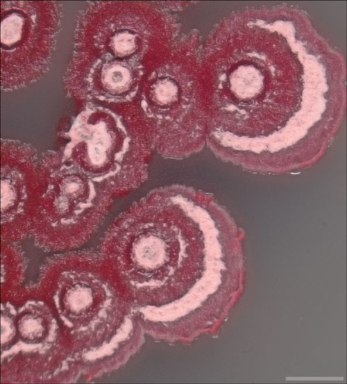
Bacteria colonies ( Streptomyces coelicolor ) in agarose, day 1 for a longitudinal, colorimetric study. Image acquired using a Leica NZ FL3 stereo microscope with a 1x lens. A SPOT RT camera and SPOT Advanced acquisition software, version 4.6 (Diagnostic Instruments, Sterling Heights, MI) were used for imaging. Color correction, sharpening, cropping, and resampling were done in Adobe Photoshop CS3 Extended, version 10.1 (Adobe Systems Incorporated, San Jose, CA). Scale bar = 10mm.
In the world of scientific research, images fall broadly into two categories: the original image and the corrected image. The original is acquired via an imaging device without any corrections applied in software. The corrected image, often referred to as enhanced, is often used for presentation and publication.
The distinction is crucial when suspicions arise about content in images. Because images are used as visual proof of experimental evidence, certain alterations to the content may be viewed as unethical. Certainly, any additions to the content from other images, or intentional alterations of visual data to accommodate the experimental hypothesis, will always be unethical. The sole means for determining the extent or the existence of alterations or additions lies in looking at the original.
A kind of reasoning can then follow: If the original is the indisputable visual evidence for experimental conclusions, only the original should be used. No alterations to visual data should be applied, and no corrections should be made for imaging device inconsistencies or shortcomings.


