Auerbach Max - An Atlas of Anatomy for Artists
Here you can read online Auerbach Max - An Atlas of Anatomy for Artists full text of the book (entire story) in english for free. Download pdf and epub, get meaning, cover and reviews about this ebook. City: New York, year: 1957;1947, publisher: Dover Publications, genre: Detective and thriller. Description of the work, (preface) as well as reviews are available. Best literature library LitArk.com created for fans of good reading and offers a wide selection of genres:
Romance novel
Science fiction
Adventure
Detective
Science
History
Home and family
Prose
Art
Politics
Computer
Non-fiction
Religion
Business
Children
Humor
Choose a favorite category and find really read worthwhile books. Enjoy immersion in the world of imagination, feel the emotions of the characters or learn something new for yourself, make an fascinating discovery.
- Book:An Atlas of Anatomy for Artists
- Author:
- Publisher:Dover Publications
- Genre:
- Year:1957;1947
- City:New York
- Rating:3 / 5
- Favourites:Add to favourites
- Your mark:
An Atlas of Anatomy for Artists: summary, description and annotation
We offer to read an annotation, description, summary or preface (depends on what the author of the book "An Atlas of Anatomy for Artists" wrote himself). If you haven't found the necessary information about the book — write in the comments, we will try to find it.
Auerbach Max: author's other books
Who wrote An Atlas of Anatomy for Artists? Find out the surname, the name of the author of the book and a list of all author's works by series.


An Atlas of Anatomy for Artists — read online for free the complete book (whole text) full work
Below is the text of the book, divided by pages. System saving the place of the last page read, allows you to conveniently read the book "An Atlas of Anatomy for Artists" online for free, without having to search again every time where you left off. Put a bookmark, and you can go to the page where you finished reading at any time.
Font size:
Interval:
Bookmark:
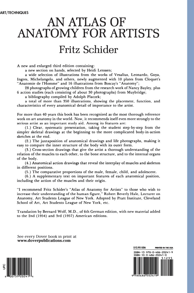
FRITZ SCHIDER
ANATOMY
FOR ARTISTS
AND TRANSLATED BY BERNARD WOLF, M.D. NEW BIBLIOGRAPHY BY ADOLF K. PLACZEK,
COLUMBIA UNIVERSITY ADDITIONAL ILLUSTRATIONS FROM THE
OLD MASTERS AND HISTORICAL SOURCES WITH A NEW SECTION ON HANDS SELECTED BY
HEIDI LENSSEN THIRD AMERICAN EDITION DOVER PUBLICATIONS, INC.
NEW YORK Copyright 1947, 1954, 1957 by Dover Publications, Inc. All rights reserved. Aufl. published by E. A. Seeman. Library of Congress Catalog Card Number: 58-3622International Standard Book NumberISBN-13: 978-0-486-20241-9
ISBN-10: 0-486-20241-0 Manufactured in the United States by Courier Corporation
20241043
www.doverpublications.com
The book has been expanded by the addition of the following material: (1) A new bibliography. (2) A wide selection of illustrations from historical sources: Vesalius, Leonardo, Goya, Degas, and others. (3) Photographic illustrations of interest to the artist which are reproduced for the first time in this book: the Nancy Bayley photographs of growing children and the Muybridge action studies. Although Schider has always been a valuable book for the study of anatomy, it is hoped that the added sections will encourage the student to study life drawing from the rich repository of material that is readily available in the great libraries and museums of the world. Rimmer and Muybridge, for example, were great teachers and students of the human figure during the nineteenth century; yet, their books are out of print at the present time. If this book introduces to the student such works as these and encourages him to investigate the artistic and photographic resources that are available, much of the purpose of the book will have been achieved.
Schider has been particularly useful in that he has never encouraged the student to follow any style other than his own. He has concentrated primarily on presenting the essential facts of anatomy in a straightforward manner leaving the student in less danger of imitating particular styles or mannerisms. This aspect of the book has not been altered; rather, the introduction of the historical material should make the student continuously aware of the variety of style and approach that is possible. 1954 Dover Publications, Inc.
NOTE: The female skeleton is clearly differentiated from the male by the small face and skull, the narrow, short thorax, and particularly the more rounded pelvis (compare the drawings).
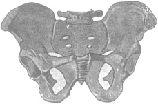 Male Pelvis demonstrates the groove between the two tuberosities at the upper end of the humerus, a typical bone groove, and the oval rough area of the humerus (insertion of the deltoid muscle). demonstrates the linea aspera, the rugh line on the posterior aspect of the femur (origin and insertion of thigh muscles), a typical bone ridge; the head of the femur, the upper cartilage-covered end of the femur, with the femoral neck and the two femoral trochanters. demonstrates the crest of the tibia, the upper portion of the S-shaped edge of the tibia, a typical bone edge. shows a tubular bone sawn across with its marrow cavity.
 The Types of Joints.
The ligaments between the femur and pelvis. The ball and socket joint consists of a spherical head which fits into a cavity, the acetabulum, and which allows motion in all directions. Flexion, extension, adduction, and circumduction are possible in this type of joint. B. Hinge Joints. .
The joints of the fingers, the inter-phalangeal joints, are shown as examples of this type. In a hinge joint, one bone has a transverse convex cylindrical surface and the other bone shows the reciprocal contour. Only flexion and extension are possible in such a joint. 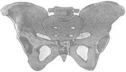 Female Pelvis C. Combination Type of Joint. of the forearm and hand is the position assumed after maximum inward rotationthe palm then faces outwards.
Supination refers to the opposite motion, i.e. rotating the palm outwards away from the body; the supinated position is the position assumed after maximum outward rotationthe palm faces forward and slightly outwards.) D. Immobile Type of Joint. . The joints between the individual wrist (carpal) and ankle (tarsal) bones and between the carpal and metacarpal, tarsal and metatarsal bones are examples of this type.
Schematic Cross-section Through a Joint.
The two mandibular fossae in which the articular processes of the mandible move. C. The occipital protuberance to which the ligamentum nuchae (ligament of the neck) isattached. D. The mastoid processes, the styloid processes, and the external occipital crest which serve for the origin or insertion of muscles. E.
The foramen magnum is the connection between the cranial cavity and the vertebral (spinal) canal. . In this drawing, significant features as far as external appearance is concerned are: A. The two frontal prominences rounded protuberances more definitely marked in children and women than in men; B. The two superciliary arches slender ridges above the orbits more distinctly marked in men than in women or children; C. The glabella a small, flat surface between the superciliary arches; D.
The temporal lines characteristically individual lines which form the lateral margins of the forehead; E. The nasal bones; F. The zygomatic bones with their very prominent zygomatic processes forming the anterior portions of the zygomatic arches; G. The chin formed by the central part of the mandible. show the skull of the newborn, viewed from above and from the left side. Sutural lines have not formed as yet.
Instead, membrane-covered spaces are present between bones concerned. The frontal bone consists of two portions, unfused as yet. demonstrates the senile skull. As a result of the teeth falling out, the mandible is thinned, the angle of the mandible obtuse, the mandible extends beyond the maxilla, and the chin protrudes. demonstrate the contours of three different skulls with their sutures.
Font size:
Interval:
Bookmark:
Similar books «An Atlas of Anatomy for Artists»
Look at similar books to An Atlas of Anatomy for Artists. We have selected literature similar in name and meaning in the hope of providing readers with more options to find new, interesting, not yet read works.
Discussion, reviews of the book An Atlas of Anatomy for Artists and just readers' own opinions. Leave your comments, write what you think about the work, its meaning or the main characters. Specify what exactly you liked and what you didn't like, and why you think so.