• Complain

Fliss Dan M. - Atlas of Surgical Approaches to Paranasal Sinuses and the Skull Base

Here you can read online Fliss Dan M. - Atlas of Surgical Approaches to Paranasal Sinuses and the Skull Base full text of the book (entire story) in english for free. Download pdf and epub, get meaning, cover and reviews about this ebook. City: Berlin;Heidelberg, year: 2016;2018, publisher: Springer, genre: Detective and thriller. Description of the work, (preface) as well as reviews are available. Best literature library LitArk.com created for fans of good reading and offers a wide selection of genres:

Romance novel Science fiction Adventure Detective Science History Home and family Prose Art Politics Computer Non-fiction Religion Business Children Humor

Choose a favorite category and find really read worthwhile books. Enjoy immersion in the world of imagination, feel the emotions of the characters or learn something new for yourself, make an fascinating discovery.

Fliss Dan M. Atlas of Surgical Approaches to Paranasal Sinuses and the Skull Base
  • Book:
    Atlas of Surgical Approaches to Paranasal Sinuses and the Skull Base
  • Author:
  • Publisher:
    Springer
  • Genre:
  • Year:
    2016;2018
  • City:
    Berlin;Heidelberg
  • Rating:
    4 / 5
  • Favourites:
    Add to favourites
  • Your mark:
    • 80
    • 1
    • 2
    • 3
    • 4
    • 5

Atlas of Surgical Approaches to Paranasal Sinuses and the Skull Base: summary, description and annotation

We offer to read an annotation, description, summary or preface (depends on what the author of the book "Atlas of Surgical Approaches to Paranasal Sinuses and the Skull Base" wrote himself). If you haven't found the necessary information about the book — write in the comments, we will try to find it.

Fliss Dan M.: author's other books


Who wrote Atlas of Surgical Approaches to Paranasal Sinuses and the Skull Base? Find out the surname, the name of the author of the book and a list of all author's works by series.

Atlas of Surgical Approaches to Paranasal Sinuses and the Skull Base — read online for free the complete book (whole text) full work

Below is the text of the book, divided by pages. System saving the place of the last page read, allows you to conveniently read the book "Atlas of Surgical Approaches to Paranasal Sinuses and the Skull Base" online for free, without having to search again every time where you left off. Put a bookmark, and you can go to the page where you finished reading at any time.

Light

Font size:

Reset

Interval:

Bookmark:

Make
Springer Berlin Heidelberg 2016
Dan M. Fliss and Ziv Gil Atlas of Surgical Approaches to Paranasal Sinuses and the Skull Base 10.1007/978-3-662-48632-0_1
1. The Cranial Base
Dan M. Fliss 1 and Ziv Gil 2
(1)
Division of Otolaryngology Head and Neck Surgery and Maxillofacial Surgery, Tel Aviv Sourasky Medical Center, Tel Aviv, Israel
(2)
The Head and Neck Center Department of Otolaryngology Head and Neck Surgery, Rambam Healthcare Campus, Haifa, Israel
Keywords
Cerebellopontine angle Cranial base Craniovertebral junction Cavernous sinus Orbit Paranasal sinuses Posterior fossa Sellar region Temporal bone
1.1 Overview
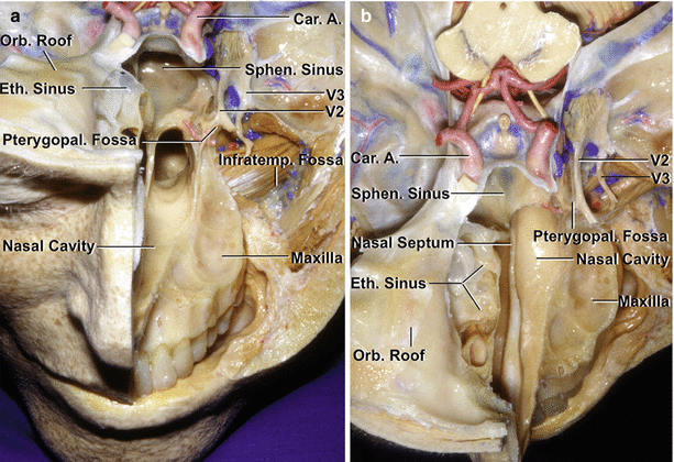
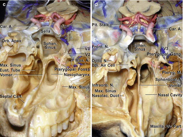
No part of the cranial base is immune to surgical pathology or to its use as a pathway to access lesions in the intra- or extracranial spaces. Tumors and multiple other lesions can involve any of the intracranial fossae and can appear in the paranasal sinuses, nasal cavity, infratemporal and pterygopalatine fossae, and orbit and in the retropharyngeal and craniocervical regions (Fig. ).
Fig 11 Anterior and middle cranial base a On the left side the floor of - photo 1Fig 11 Anterior and middle cranial base a On the left side the floor of - photo 2
Fig. 1.1
Anterior and middle cranial base. ( a ) On the left side, the floor of the anterior fossa and the upper portion of the maxilla have been removed to expose the structures deep to the anterior and middle cranial fossa. The frontal, ethmoidal, and sphenoid sinuses and the nasal cavity lie below the medial part of the anterior cranial base. The orbit and maxilla are located below the lateral part of the anterior cranial base. The sphenoid sinus and sella are located in the medial part of the middle cranial base, and the infratemporal and pterygopalatine fossae are located below the lateral part of the middle cranial base. The carotid arteries pass upward on the medial part of the middle cranial base and are intimately related to the sphenoid and cavernous sinuses. The infratemporal fossa, which contains branches of the mandibular nerve, pterygoid muscles, pterygoid venous plexus, and maxillary artery, is located below the middle cranial base and greater sphenoid wing. The alveolar process of the maxilla, which encloses the roots of the upper teeth, has been preserved on the left side. The maxillary nerve enters the pterygopalatine fossa, which is located medial to the infratemporal fossa between the posterior wall of the maxilla and the pterygoid process of the sphenoid bone. ( b ) Superior view of the anterior and middle cranial base. The infratemporal fossa is located posterolateral to the maxilla. The right ethmoid air cells are exposed on the medial side of the right orbit. The nasal cavity extends upward between the ethmoid sinuses. ( c ) Oblique anterior view. The facial structures on the right side have been removed to expose the orbital apex located above the maxillary sinus. The wall of the right maxillary sinus forms the floor of the orbit, much of the lateral wall of the nasal cavity, and the anterior wall of the pterygopalatine and infratemporal fossa. On the left side, the mandibular nerve enters the infratemporal fossa. The maxillary nerve enters the pterygopalatine fossa, which is located in the lateral wall of the nasal cavity and contains the maxillary nerve, pterygopalatine ganglion, and terminal branches of the maxillary artery. ( d ) Anterior view. The orbital apex is located above the pterygopalatine fossa. The frontal branch of the ophthalmic nerve passes along the roof of the orbit, and the infraorbital branch of the maxillary nerve courses in the floor of the orbit. The posterior ethmoid air cells are located medial to the orbital apex. The vomer forms the posterior part of the nasal septum and attaches to the maxilla and palatine bones below and to the body of the sphenoid bone above. The sphenoid sinus is located in the middle cranial base below the sella turcica. The upper brainstem is seen in the posterior part of the exposure. A artery, Alv alveolar, Car carotid, Cart cartilage, CN cranial nerve, Eth ethmoid, Eust eustachian, Front frontal, Gang ganglion, Infraorb infraorbital, Infratemp infratemporal, M muscle, Max maxillary, N nerve, Nasolac nasolacrimal, Orb orbital, Pit pituitary, Pteryg pterygoid, Pterygopal pterygopalatine, Sphen Sphenoid, Sphenopal sphenopalatine (From Rhoton Jr AL. The anterior and middle cranial base. Neurosurgery. 2002;51(1 Suppl):S273302; with permission )
Fig 12 Specimen divided in the midsagittal plane to show the endoscopic - photo 3
Fig. 1.2
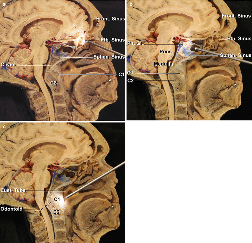
Specimen divided in the midsagittal plane to show the endoscopic endonasal approaches to the cranial base in midline through the frontal sinus, ethmoid sinus, sphenoid sinus, and nasopharynx. ( a ) Anterior cranial base approaches in midline through the frontal and ethmoid sinuses. ( b ) Approaches through the sphenoid sinus to the sellar region. ( c ) The endoscope is oriented downward to the nasopharynx for C1C2 approaches. Eth ethmoid, Eust eustachian, Front frontal, Gl gland, Pit pituitary, Sphen sphenoid
The skull is divided into the cranium and the facial skeleton. The cranium is divided into the calvarium and the cranial base. The cranial base has an endocranial surface, which faces the brain, and an exocranial surface, which faces the nasal cavity and sinuses, orbits, pharynx, infratemporal and pterygopalatine fossae, and the parapharyngeal and intrapetrosal spaces (Fig. ). On the endocranial side, the border between the anterior and middle cranial bases is the sphenoid ridge, which is joined medially by the chiasmatic sulcus, and the border between the middle and posterior cranial bases is the petrous ridges, which are joined by the dorsum sellae and posterior clinoid processes. On the exocranial side, the anterior and middle cranial bases are divided at the level of a transverse line extending through the pterygomaxillary fissures and the pterygopalatine fossae at the upper level and the posterior edge of the alveolar process of the maxilla at the lower level. Medially, this corresponds to the anterior part of the attachment of the vomer to the sphenoid bone. The middle and posterior cranial bases are separated by a transverse line crossing at or near the posterior border of the vomer-sphenoid junction, the foramen lacerum, carotid canal, jugular foramen, styloid process, and the mastoid tip.
Fig 13 Lateral view of the anterior middle and posterior cranial base a - photo 4
Fig. 1.3
Lateral view of the anterior, middle, and posterior cranial base. ( a ) The bone and structures lateral to the orbit, infratemporal, and pterygopalatine fossa and the parapharyngeal space and petrous part of the temporal bone have been removed to expose the structures below the anterior, middle, and posterior cranial base. The orbit and maxillary sinus are located below the anterior cranial base. The infratemporal and pterygopalatine fossae and the parapharyngeal space are located below the middle cranial base, and the suboccipital area is located below the temporal and occipital bones. The first trigeminal division is related to the upper part of the orbit. The second trigeminal branch is related to the lower part of the orbit and maxilla. The mandibular nerve exits the cranium through the foramen ovale and enters the infratemporal fossa. The pterygoid and levator and tensor veli palatini muscles have been removed to expose the eustachian tube and its opening into the nasal pharynx. The lateral part of the temporal bone has been removed to expose the cochlea, vestibule, and semicircular canals. The petrous carotid passes upward and turns medially below the cochlea. The sigmoid sinus turns downward under the semicircular canals and vestibule where the jugular bulb is located. The segment of the vertebral artery passing behind the atlanto-occipital joint is located below the posterior cranial base. ( b ) The dura has been opened to show the relationships of the frontal and temporal lobes and the cerebellum to the cranial base. The orbit is exposed below the frontal lobe. The pterygopalatine and infratemporal fossae and the temporal bone are located below the temporal lobe. The jugular bulb and internal jugular vein have been removed to show cranial nerves 9 through 12 exiting the jugular foramen. A artery, Car carotid, CN cranial nerve, Eust eustachian, Front frontal, Gr greater, Inf inferior, Infraorb infraorbital, Infratemp infratemporal, Int internal, Jug jugular, Lat lateral, M muscle, Max maxillary, N nerve, Ped peduncle, Pet petrosal, Pterygopal pterygopalatine, Rec rectus, Semicirc semicircular, Sphen sphenoid, Temp temporal, V vein, Vert vertebral (From Rhoton Jr AL. The anterior and middle cranial base. Neurosurgery. 2002;51(1 Suppl):S273302.; with permission )
Next page
Light

Font size:

Reset

Interval:

Bookmark:

Make

Similar books «Atlas of Surgical Approaches to Paranasal Sinuses and the Skull Base»

Look at similar books to Atlas of Surgical Approaches to Paranasal Sinuses and the Skull Base. We have selected literature similar in name and meaning in the hope of providing readers with more options to find new, interesting, not yet read works.


Reviews about «Atlas of Surgical Approaches to Paranasal Sinuses and the Skull Base»

Discussion, reviews of the book Atlas of Surgical Approaches to Paranasal Sinuses and the Skull Base and just readers' own opinions. Leave your comments, write what you think about the work, its meaning or the main characters. Specify what exactly you liked and what you didn't like, and why you think so.