Andreas Raabe - The Craniotomy Atlas
Here you can read online Andreas Raabe - The Craniotomy Atlas full text of the book (entire story) in english for free. Download pdf and epub, get meaning, cover and reviews about this ebook. year: 2019, publisher: Thieme, genre: Home and family. Description of the work, (preface) as well as reviews are available. Best literature library LitArk.com created for fans of good reading and offers a wide selection of genres:
Romance novel
Science fiction
Adventure
Detective
Science
History
Home and family
Prose
Art
Politics
Computer
Non-fiction
Religion
Business
Children
Humor
Choose a favorite category and find really read worthwhile books. Enjoy immersion in the world of imagination, feel the emotions of the characters or learn something new for yourself, make an fascinating discovery.

- Book:The Craniotomy Atlas
- Author:
- Publisher:Thieme
- Genre:
- Year:2019
- Rating:4 / 5
- Favourites:Add to favourites
- Your mark:
The Craniotomy Atlas: summary, description and annotation
We offer to read an annotation, description, summary or preface (depends on what the author of the book "The Craniotomy Atlas" wrote himself). If you haven't found the necessary information about the book — write in the comments, we will try to find it.
Andreas Raabe: author's other books
Who wrote The Craniotomy Atlas? Find out the surname, the name of the author of the book and a list of all author's works by series.


The Craniotomy Atlas — read online for free the complete book (whole text) full work
Below is the text of the book, divided by pages. System saving the place of the last page read, allows you to conveniently read the book "The Craniotomy Atlas" online for free, without having to search again every time where you left off. Put a bookmark, and you can go to the page where you finished reading at any time.
Font size:
Interval:
Bookmark:
I would like to express my deep gratitude to Anja Giger and Alain Blank, who provided the superb illustrations for this book. Over a period of 3 years, it was always a pleasure to sit together and discuss the details of the authors photographs and how these should be depicted in the illustrations. Without their artistic skills and their invaluable contribution, this book would not have been possible.
I am specifically grateful to Luisa Tonarelli, who accompanied the development of this book from the very first chapter to the final printed version. Her help, advice, expertise, and hard work were indispensable in bringing this volume to publication.
Finally, I would like to thank Susan Kaplan, Irena Zubak, Janine Abu-Isa, Katharina Lutz, Michael Murek, David Bervini, Johannes Goldberg, Levin Hni, and Jonathan Rychen for their time and advice during the review of the chapters of this book.
Andreas Raabe
Andreas Raabe and Peter A. Winkler
There are four basic categories of supratentorial and infratentorial craniotomy:
Convexity craniotomies may be performed anywhere according to the surgical target and goal of the operation. They range from burr holes and mini-craniotomy to decompressive hemicraniectomy, which is the most extensive variant.
Midline craniotomies are used for midline approaches that take advantage of subdural anatomical corridors to reach superficial, deep, or contralateral targets. The supratentorial suboccipital craniotomy with an intradural approach along the falx and the tentorium or an infratentorial suboccipital craniotomy with a supracerebellar approach are possible variants.
Skull base craniotomies range from the frontal midline to the foramen magnum, covering the entire skull base. demonstrate the continuum of approaches which are often overlapping and are named according to their location at the skull base.
Skull base extensions are added to standard skull base craniotomies. They allow access with angles of approach or to structures that cannot be easily reached with standard skull base craniotomies. Typical skull base extensions are anterior clinoidectomy, removal of the orbital rim or zygoma (orbitozygomatic), transpetrosal approaches, the suprameatal extension after retrosigmoid craniotomy or the far-(enough) lateral extension to the foramen magnum (see , Skull Base Extensions).
Supratentorial skull base craniotomies can be divided according to their location, their frontal and temporal extension (size), and their relation to the sylvian fissure. There is no uniform classification, but the following general rules may serve as a guide to the terminology (see ).
Table 1.1 Systematics of skull base craniotomiessupratentorial
Location | Description |
Median frontobasal | Mostly bilateral. Target: medial frontal base, anterior midline. |
Frontolateral | Extends 13 cm lateral to the midline to approximately the sphenoid wing, but does not cross it. The proximal sylvian fissure is exposed intradurally, and targets within the sylvian fissure, the anterior skull base, and the temporal lobe can be reached. There are mini- and standard sizes. Frontolateral is the term that was historically first used for this approach. |
Supraorbital | Usually a smaller variant of the frontolateral approach; typically by eyebrow (transciliary) incision, which limits the size of the craniotomy. Extends 2.53 cm lateral to the midline to approximately the sphenoid wing, but does not cross it. The proximal sylvian fissure is exposed intradurally, and targets in the sylvian fissure, skull base, and temporal lobe can be reached. Some surgeons use the term supraorbital as synonymous with frontolateral. |
Pterional | Extends 13 cm lateral to the midline to the anterior temporal region: centered around the H of the sutures that form the pterion (see , Craniocerebral Topography). The sphenoid wing is always crossed. Typically defined as two-thirds of the craniotomy frontal and one-third temporal exposure of variable sizes (2:1). There is also a mini-pterional variant. |
Frontotemporal | Usually a large exposure (1:1 to 2:1 frontal:temporal) centered above the sphenoid wing = sylvian fissure. |
Anterior temporal | Sphenoid wing is crossed. |
Temporobasal | The exact position varies according to the surgical target: does not cross the sphenoid wing. Typically used for subtemporal intradural approaches. There may be a more anterior and a more posterior variant. |
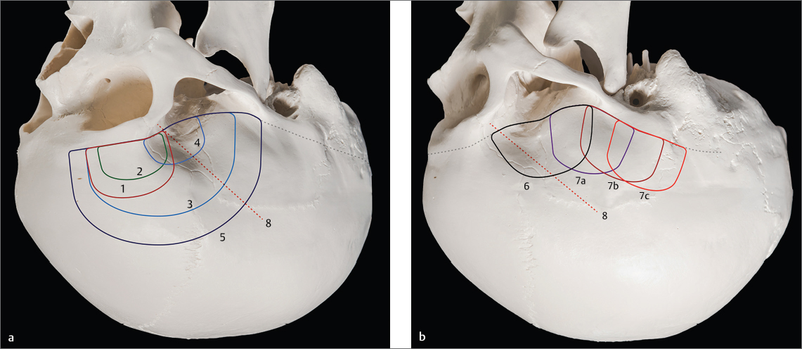
Fig. 1.1 Systematics of skull base craniotomiessupratentorial. Supratentorial frontotemporal skull base craniotomies, 45 view (a) and lateral view (b). 1, frontolateral; 2, supraorbital; 3, standard pterional; 4, mini-pterional; 5, frontotemporal; 6, anterior temporal; 7ac anterior, middle, posterior temporobasal; 8, sylvian fissure/sphenoid wing.
for further details).
Table 1.2 Systematics of skull base craniotomiesinfratentorial
Location | Description |
Suboccipital median infra-transverse-sinus | Midline craniotomy for supracerebellar median or paramedian approaches, e.g., for access to the pineal region or tentorial dural fistulas. |
Suboccipital lateral infra-transverse-sinus | These are craniotomies based on the same principle as the midline craniotomies for an intradural approach along the subdural space parallel to the tentorium. Typically, they are used for supracerebellar lateral approaches to the midbrain or other regions. They are horizontally oriented compared to the retrosigmoid craniotomy, with more exposure along the transverse sinus and less along the sigmoid sinus. A modification is the suboccipital far-lateral infra-transverse-sinus craniotomy. |
Retrosigmoid | Typically ranges from the transverse sinus to the base of the posterior fossa along the sigmoid sinus to gain access to the cerebellopontine angle. May vary in size and be centered more superiorly or inferiorly: vertically oriented. |
Suboccipital median periforaminal craniotomy with opening of the foramen magnum | Typically bilateral, there is a mini-version, for example, in Chiari-decompression surgery. |
Suboccipital lateral periforaminal craniotomy with opening of the foramen magnum | The lateral suboccipital craniotomy with opening of the foramen magnum is the basic craniotomy for the far lateral approach which can be regarded as a skull base extension of the basal suboccipital craniotomy. |
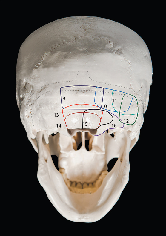
Fig. 1.2 Systematics of skull base craniotomiesinfratentorial. Craniotomies of the posterior fossa. 9, suboccipital median infra-transverse-sinus; 10, suboccipital lateral infra-transverse-sinus; 11, suboccipital far-lateral infra-transverse-sinus; 12, retrosigmoid; 13, suboccipital median periforaminal (with opening of the foramen magnum); 14, mini-suboccipital median periforaminal (with opening of the foramen magnum); 15, suboccipital lateral periforaminal (with opening of the foramen magnum); 16, far-lateral extension.
Font size:
Interval:
Bookmark:
Similar books «The Craniotomy Atlas»
Look at similar books to The Craniotomy Atlas. We have selected literature similar in name and meaning in the hope of providing readers with more options to find new, interesting, not yet read works.
Discussion, reviews of the book The Craniotomy Atlas and just readers' own opinions. Leave your comments, write what you think about the work, its meaning or the main characters. Specify what exactly you liked and what you didn't like, and why you think so.