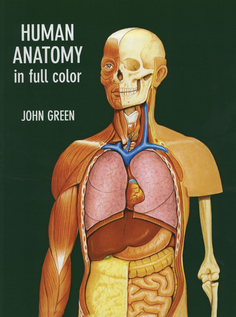
Human Anatomy
in Full Color
Illustrations by John Green
Text by Dr. John W. Harcup
S.B.St.J., M.R.C.G.P., M.R.C.S., D.Obst. R.C.O.G. Fellow of the Royal Society of Medicine
DOVER PUBLICATIONS, INC. MINEOLA, NEW YORK Copyright
Illustrations copyright 1996 by John Green.
Text copyright 1996 by John W. Harcup. All rights reserved.
Bibliographical Note
Human Anatomy in Full Color is a new work, first published by Dover Publications, Inc., in 1996.
Library of Congress Cataloging-in-Publication Data
Green, John, 1948 Human anatomy in full color / illustrations by John Green ; text by John W. p. cm. cm.
Includes index. ISBN 0-486-29065-4 (pbk.) 1. Human anatomyAtlases. I. Harcup, John W. Title. Title.
QM25.G74 1996 611.00222dc20 96-42325 CIP
Manufactured in the United States by Courier Corporation 29065410 www.doverpublications.com
INTRODUCTION
The human body is an amazing structure, so complex that, even today, not all its functions are fully understood. Certain cells are directed by genes to divide into organs with specialized functions. The sensessight, hearing, taste, touch and smellconnect us to the outside world. The body is capable of detecting minute changes in the environment. It can adapt to different conditions and extremes of heat and cold. Quickly, it can prepare for flight or fight to protect itself from danger.
However, when the enemy is within, as tiny as a bacterium or virus, the human body has its own protective mechanism in the form of white blood cells and antibodies that kill the invader. Many ingested poisons are made harmless in the liver; waste products are efficiently excreted by the kidneys and alimentary canal. The brain can appreciate beauty, devise art forms, compose music and create pictures, solve puzzles, design clothes, invent rockets to probe space and program computers. With specialized appendagesthe handsthe body can make models and sculptures, paint paintings, knit, embroider exquisite garments, communicate by writing and drive cars and planes with incredible accuracy. The legs provide a means of locomotion. The ability to walk upright allows the arms freedom.
John Green has drawn the human body to make it clearly understandable. In the text, I have tried to explain the body and to answer some of the questions I have been asked, as a doctor, on this subject.
John W. Harcup Malvern, England 1996
CONTENTS
THE SKELETON
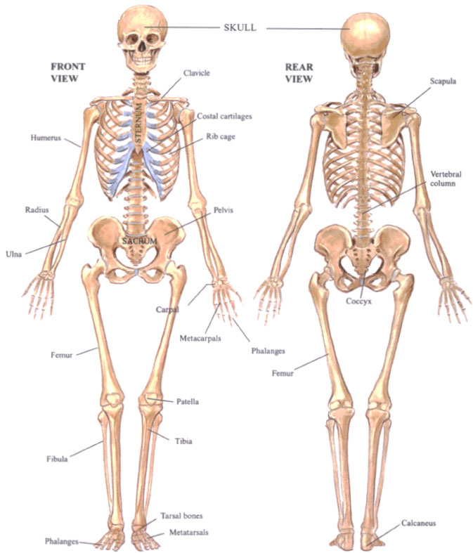
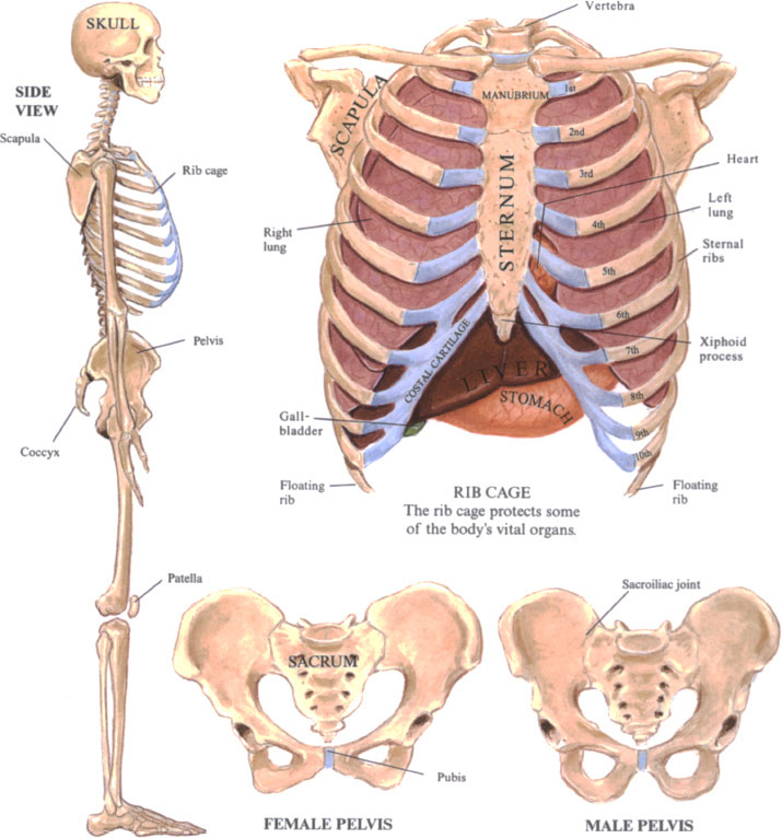
The 206 bones in the skeleton form the framework of the body, accounting for one seventh of an adult males weight. The 80 bones of the head, face, neck and trunk are called the axial skeleton. The appendicular skeleton, comprising the limbs or appendages, is made up of 32 bones in each adult upper limb and 31 in each lower limb.
Sesamoid bones are connected to a tendon rather than to another bone; everybody has at least twothe patella (kneecap) of each leg but some people have them in other sites such as the foot or over the first joint in the thumb. (If present, these are extra bones and are always small.) The largest bone in the body is the femur, the smallest are the ossicles in the middle ear. Bones have several important functions. They are the support system of the body for soft tissues and anchorage points for muscles. To allow movement of the body, individual bones must be able to move in relationship to one another. This happens at joints, where the bones articulate with each other, the surfaces that touch being termed articular surfaces.
Groups of bones form protective cages: The skull protects the brain, and the lungs and heart are enclosed by the bones of the vertebral column (spine), ribs and sternum (breastbone). There are 12 pairs of ribs; the lower two pairs of ribs float and are not connected to the gristle or cartilage that composes the junction between the ribs and breastbone. In the pelvis the bone arrangement protects vital organs of the lower abdomen, but the female pelvis is wider than the male to allow space for a baby to pass through when it is born. The sacrum ends in the coccyx, which is a residual tail.
Inside bones, red and white blood cells and platelets are manufactured and essential elements such as calcium and phosphate are stored.
BONES & JOINTS
There are three kinds of bonelong (e.g., thigh and arm); flat (the shoulders, skull, ribs and pelvis) and irregular (the spinal column, ankle and wrist, although the last are sometimes called short bones).
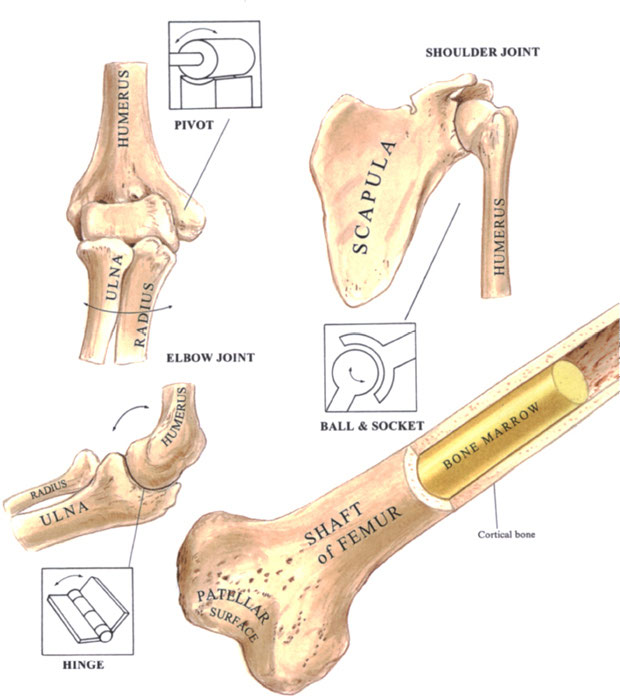
Bones are covered by a tough sheath, the periosteum, which contains many small nerve endings. This is why a blow on a bone such as the shin is so painful. The two types of bone, spongy (cancellous) and cortical, are composed of Haversian canals carrying nerves, blood vessels and lymphatics, surrounded by plates of bone and spaces containing bone cellsosteoblasts and osteocytes, which produce bone, and osteoclasts, which reabsorb it. Even in adults, 15 percent of bone may be remodeled in a year. Spongy bone has the appearance of a sponge as the canals are larger and the spaces contain bone marrow, comprised of blood and fat cells. In long bones, red blood cells are manufactured in the red bone marrow, which predominates in childhood, but in adults, this is replaced mainly by yellow, fatty marrow in the medullary cavity.
Bones are formed from calcium and phosphorus derived from diet and combined to form calcium phosphate. Long bones grow as new bone is deposited by the layer beneath the periosteum and at bands of cartilage, called epiphyseal plates, near the articular surfaces, until full growth is reached between the ages of 18 and 25, when bone replaces this cartilage. The age at which a bone stops growing varies from one bone to another. A joint is formed where two bones meet. The surfaces on which they move on one another are articular surfaces. They are covered in articular cartilage, which acts as a cushion.
There are three types of joints. Fibrous joints are fixed and immovable, such as the bones of the skull. Cartilaginous joints are slightly movable joints in which a plate of fibrocartilage separates the bones, as in the breastbone (sternum) and the vertebral bodies of the spinal column. Synovial joints are freely movable and lined by a membrane producing a lubricant, synovial fluid. There are seven varieties of synovial joint. A hinge joint, like the elbow, moves up and down like the lid of a box.
A pivot joint allows rotation only, as in the radius and ulna bones at the elbow, or the second bone in the neck. In a plane joint the surfaces are flat, as in the wrist and foot. Ball and socket joints, such as the shoulder and hip, can move in all directions. Condyloid joints are modified ball-and-socket joints that cannot move upward, such as the fingers and knuckle joints. An ellipsoid joint is another modified ball-and-socket joint in the wrist involving the carpal and radius bones, capable of all movements except rotation. A saddle joint, such as the thumb, moves on two axes at right angles to each other.


