This edition published in 2014 by:
The Rosen Publishing Group, Inc.
29 East 21st Street
New York, NY 10010
Additional end matter copyright 2014 by The Rosen Publishing Group, Inc.
All rights reserved. No part of this book may be reproduced in any form without permission in writing from the publisher, except by a reviewer.
Library of Congress Cataloging-in-Publication Data
Wanjie, Anne.
The basics of cell biology/Anne Wanjie.1st ed.New York:
Rosen, 2014
p. cm.(Core concepts)
Includes bibliographical references and index.
ISBN 978-1-4777-0548-3 (library binding)
1. CytologyJuvenile literature. I. Title.
QH582.5 W36 2014
571.6
Manufactured in the United States of America
CPSIA Compliance Information: Batch #S13YA: For further information, contact Rosen Publishing, New York, New York, at 1-800-237-9932.
2004 Brown Bear Books Ltd.
CONTENTS
CHAPTER 1: AN INTRODUCTION TO THE CELL
CHAPTER 2: A VARIETY OF TYPES
CHAPTER 3: CELL LOCOMOTION AND SUPPORT
CHAPTER 4: A LOOK INSIDE THE CELL
CHAPTER 5: HOW CELLS COMMUNICATE
CHAPTER 6: UNDERSTANDING THE CELL CYCLE
CHAPTER 7: ABNORMAL REPRODUCTION
CHAPTER 8: BIOGRAPHIES: FRANCIS CRICK, ROSALIND FRANKLIN, AND JAMES WATSON
GLOSSARY
FOR MORE INFORMATION
FOR FURTHER READING
INDEX
CHAPTER ONE
AN INTRODUCTION TO THE CELL
Cells are the building blocks of life. Your body contains trillions of cells, but many creatures are just a single cell.
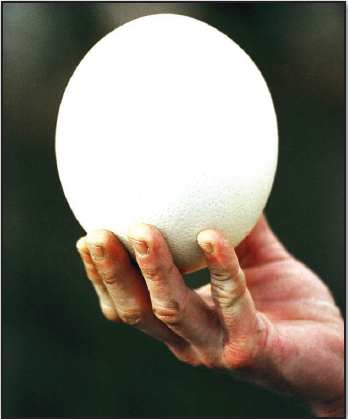
M any areas of biology depend on an understanding of cells. The largest single cell, an ostrich egg yolk, is the size of a baseball, but cells can be so tiny that a hundred placed in a row would fit on the period at the end of this sentence. Tiny cells such as protists and bacteria are usually measured in nanometers. A nanometer is 1 millionth of a millimeter. Most plants and animals are made up of billions or even trillions of cells. The study of cells is called cytology.
Cells work together to carry out important functions within an organism. Many cells are specialized to carry out certain tasks. Red blood cells, for example, are specialized for transporting oxygen around the body.
Organisms like bacteria consist of just single cells. Each cell carries out all the processes it needs to survive.
Ostrich eggs contain the largest single cells on Earth. Each cell is the yolk cell that provides food for the growing ostrich chick. However, the yolk cells inside the eggs of many dinosaurs and some giant extinct birds such as moas were probably much larger.
THE CELL LIFE CYCLE
The cells that make up your body go through a life cycle. It consists of a period of growth followed by division to produce a new pair of cells. During the period of growth cells produce chemicals, make energy from food, and provide structural support for the organism. Communication between the different parts of a cell and between a cell and its neighbors is very important to ensure they work together for the benefit of the organism.
Cells contain structures such as the cell membrane, cell wall (in plants and bacteria only), and a series of fibers called the cytoskeleton. They support the cell. Movement often depends on hairlike extensions of the cell. Long single extensions are called flagella. Groups of shorter projections that beat rhythmically are called cilia.
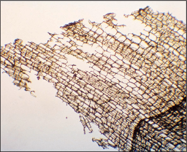
A slice of cork under a light microscope. Robert Hooke would have seen something similar as he peered through his microscope in 1667.
VIEWING CELLS UNDER A MICROSCOPE
The first microscope was made by a Dutch eyeglass-maker, Zacharias Janssen (1580c.1638). Later scientists improved on this early version. Englishman Robert Hooke (16351703) used his microscope to look closely at a thin slice of cork. Hooke used the word cell to describe the units he could see, since they reminded him of the small rooms called cells that monks lived in.
Hooke believed the cork cells were empty and that the walls were made of living material. Improvements in microscope design enabled Dutch scientist Anton van Leeuwenhoek (1632 1723) to study the cellular world in far greater detail. Leeuwenhoek was the first person to see protists, blood cells, and sperm. He also noted the presence of some cell contents such as the chloroplasts, though he was unable to describe them accurately.
In the nineteenth century microscope technology advanced further. Cytologists (cell biologists) could fully observe cells and their contents. Study of the very finest detail became possible with the invention of the electron microscope in the 1950s.
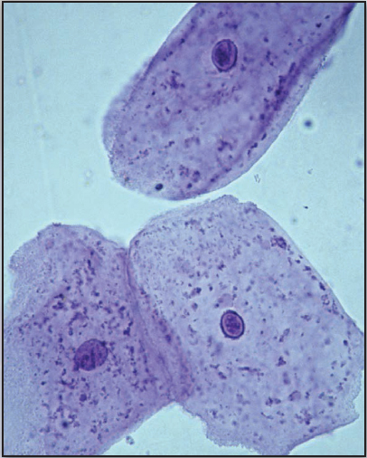
These are cheek cells. They are easy to collect from the inside of a persons cheek. Cheek cells are often the source of DNA used for DNA fingerprinting, an important tool in police work.
WORKING TOGETHER
Similar cells work together and become specialized in their functions to form tissues, such as muscles or blood in animals. A number of tissues working together make up an organ, such as a heart, stomach, or kidney. Groups of organs work together to form systems, such as the digestive system.
A NEW THEORY
Although cells were discovered and named by Robert Hooke, the idea that cells form the basic unit of living organisms was established by German biologists Matthias Schleiden (18041881) and Theodor Schwann (1810 1882). This became known as the cell theory. Schleiden studied plant cells, and Schwann studied animal cells. Later, German physician Rudolf Virchow (18211902) showed that all cells arise from preexisting cells through cell division.
PROKARYOTES AND EUKARYOTES
Cytologists divide cells into two types, prokaryotes and eukaryotes. Prokaryotes are creatures such as bacteria. They are single-celled organisms, although some occur in chains or clusters of many thousands of individuals.
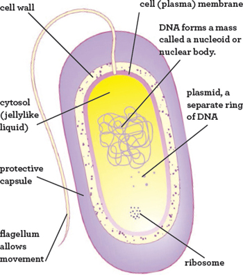
The DNA of a prokaryote cell floats freely in a region called the nucleoid. The rest of the cell is called the cytoplasm. It contains a thick, jellylike liquid called cytosol and tiny structures called ribosomes. Ribosomes use instructions encoded in the DNA to produce proteins. The cell is wrapped by a cell (plasma) membrane and, in many cases, a tough wall, too.
INSIDE A PROKARYOTE CEL
This is a bacterial cell. There is great diversity of bacterial form and function, but all have ribosomes, a cell membrane and wall, and DNA loose in a nucleoid, which is not enclosed by a membrane.
ENDOSYMBIOSIS
Scientists believe that the mitochondria that occur inside eukaryote cells descend from freeliving prokaryotes. This theory is called endosymbiosis. Evidence comes from the fact that mitochondria have their own DNA separate from the nucleus.

