Tracy I George - Atlas of Bone Marrow Pathology
Here you can read online Tracy I George - Atlas of Bone Marrow Pathology full text of the book (entire story) in english for free. Download pdf and epub, get meaning, cover and reviews about this ebook. year: 2018, publisher: Springer, genre: Home and family. Description of the work, (preface) as well as reviews are available. Best literature library LitArk.com created for fans of good reading and offers a wide selection of genres:
Romance novel
Science fiction
Adventure
Detective
Science
History
Home and family
Prose
Art
Politics
Computer
Non-fiction
Religion
Business
Children
Humor
Choose a favorite category and find really read worthwhile books. Enjoy immersion in the world of imagination, feel the emotions of the characters or learn something new for yourself, make an fascinating discovery.

- Book:Atlas of Bone Marrow Pathology
- Author:
- Publisher:Springer
- Genre:
- Year:2018
- Rating:4 / 5
- Favourites:Add to favourites
- Your mark:
Atlas of Bone Marrow Pathology: summary, description and annotation
We offer to read an annotation, description, summary or preface (depends on what the author of the book "Atlas of Bone Marrow Pathology" wrote himself). If you haven't found the necessary information about the book — write in the comments, we will try to find it.
Tracy I George: author's other books
Who wrote Atlas of Bone Marrow Pathology? Find out the surname, the name of the author of the book and a list of all author's works by series.


Atlas of Bone Marrow Pathology — read online for free the complete book (whole text) full work
Below is the text of the book, divided by pages. System saving the place of the last page read, allows you to conveniently read the book "Atlas of Bone Marrow Pathology" online for free, without having to search again every time where you left off. Put a bookmark, and you can go to the page where you finished reading at any time.
Font size:
Interval:
Bookmark:
Cell type | Characteristic morphology | Description |
|---|---|---|
Pronormoblast (proerythroblast) | 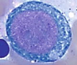 | The most immature and largest cells in erythroid lineage (1224 m), relatively high nuclear to cytoplasmic (N/C) ratio (78:1), round to slightly oval nucleus, finely reticulated chromatin, prominent nucleoli ( 1), and agranular basophilic cytoplasm |
Basophilic normoblast | 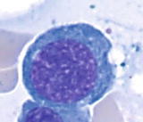 | Smaller cells (1017 m) than pronormoblast, round nucleus, high N/C ratio (6:1), open to slightly condensed chromatin, distinct parachromatin, rarely visible or absent nucleoli in later stage, and deep basophilic cytoplasm |
Polychromatophilic normoblast | 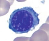 | Smaller cells (1015 m) and lower N/C ratio (4:1) than basophilic normoblasts, round nucleus with condensed chromatin, often cartwheel appearance, visible perinuclear halo, no nucleoli, and blue-gray to pink-gray cytoplasm |
Orthochromic normoblast | 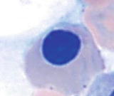 | More mature and smaller cells (812 m) than polychromatophilic normoblast, abundant cytoplasm (N/C ratio 1:2) with pink-orange and minimally basophilic color similar to erythrocytes, round nucleus, and densely condensed or pyknotic chromatin |
Erythrocyte | 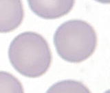 | The most mature cells (78.5 m), pink-orange to salmon color, and no nucleus |
Cell type | Characteristic morphology | Description |
|---|---|---|
Myeloblast | 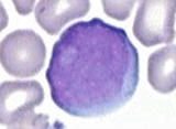 | The most immature granulocytic cells (1520 m), with high N/C ratio (47:1), round to oval nucleus, fine to reticular chromatin with distinct nucleoli (15), and moderately basophilic cytoplasm with absent or minimal azurophilic granules |
Promyelocyte | 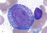 | Slightly larger cells (1424 m) than myeloblasts, with high N/C ratio (35:1), eccentric round to oval nucleus, slightly coarse or finely reticular chromatin, distinct nucleoli (13), basophilic cytoplasm with paranuclear hof and prominent azurophilic (primary) granules, which may overlie the nucleus |
Myelocyte | 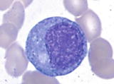 | Slightly smaller cells (1018 m) than blasts, with more abundant cytoplasm (N/C ratio 12:1), eccentric round to oval nucleus, more condensed chromatin, no nucleoli, bluish to pink cytoplasm with paranuclear hof, abundant lilac (secondary) granules, and scattered few azurophilic (primary) granules |
Metamyelocyte | 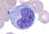 | Size similar to or slightly smaller (1018 m) than myelocytes, with abundant cytoplasm (N/C ratio 11.5:1), indented or kidney-shaped nucleus (indentation less than half the width of the nuclear margin), condensed chromatin, no nucleoli, pinkish cytoplasm with many secondary granules and rare primary granules |
Band neutrophil |
Font size:
Interval:
Bookmark:
Similar books «Atlas of Bone Marrow Pathology»
Look at similar books to Atlas of Bone Marrow Pathology. We have selected literature similar in name and meaning in the hope of providing readers with more options to find new, interesting, not yet read works.
Discussion, reviews of the book Atlas of Bone Marrow Pathology and just readers' own opinions. Leave your comments, write what you think about the work, its meaning or the main characters. Specify what exactly you liked and what you didn't like, and why you think so.